3.2. Catalytic Performance
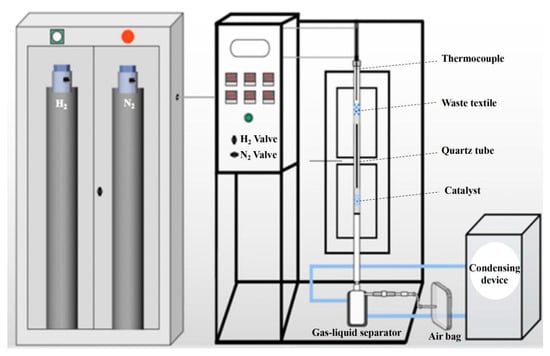
The fresh catalyst was evaluated in the pyrolysis–catalytic process; as shown in Figure 3a, the gas yield was 67.87%, the liquid yield was 14.47%, and the remaining fraction comprised a solid yield. The permanent gasses produced during the catalytic step primarily consisted of H2, CO, and CO2, with trace amounts of light hydrocarbons such as CH4, C2H4, and C2H6 also detected; the gas compositions are displayed in Figure 3b. The H2 concentration reached 35.08%, which could provide a reductive atmosphere to change the status of the Ni species [21,25]. Thus, the catalytic performances of the catalysts after hydrogen reduction at different temperatures were investigated. The gas yield increased from 67.87% to 78.46% as the hydrogen reduction temperature was raised from 300 to 800 °C, while the liquid yield decreased from 14.47% to 4.46%, suggesting the increase in the reduction temperature was beneficial to improve the permanent gasses’ yield. It might be ascribed to the production of more metallic Ni species at higher reduction temperature, which would promote C-C and C-H cleavage [26], leading to converting long-chain compounds to short-chain permanent gasses. The solid yield was almost identical at different reduction temperature, due to the formation of solid carbon tightly related to the reaction conditions of pyrolysis section rather than the catalytic section.
The impact of reduction temperature on the gas composition is shown in Figure 3b. As the reduction temperature was 300 °C, the gas compositions changed negligibly compared to the calcined catalyst. When the reduction temperature increased from 400 to 700 °C, a gradual increase in the H2 content from 35.53% to 41.50% coupled with a decrease in the CO content piecemeal from 33.02% to 27.76% was observed. However, the H2 content decreased slightly when the reduction temperature further increased from 700 to 800 °C. When the reduction temperature reached 700 °C, the CH4 concentration increased significantly, followed by the CO concentration decreased. According to the above descriptions, it was not tough to figure out the variation tendency of the CO2 content. It is widely recognized that Ni0 species serve as the active sites for CO methanation reaction on Ni-based catalysts [27]. Therefore, we could infer that a considerable amount of Ni0 species was generated after H2 reduction at a temperature of 700 °C and above, which could be further verified by characterizations.
3.3. Catalyst Characterizations
Figure 4a shows the N2 adsorption–desorption isotherms for both the fresh and reduced catalysts. Both samples exhibit type IV isotherms with H3 hysteresis loops and no saturated adsorption plateau [28], suggesting the catalysts were mesoporous materials with intergranular pores of randomly aggregated nanoparticles [29]. The hysteresis loop width in the fresh sample was almost the same as that in the reduced sample, implying both of them owned similar specific surface areas. The pore size distribution profiles, ranging from 2 to 35 nm, are shown in Figure 4b, ascribing to the mesoporous structures. As shown in Table 1, after H2 reduction at 700 °C, the specific surface area slightly decreased, the pore volume remained nearly constant, and the average pore diameter increased from 8.30 nm to 9.02 nm, which related to the collapse of pores during the reduction atmosphere. Nevertheless, these mild variations indicated that the H2 reduction addressed little impact on the structure of catalyst samples.
To investigate the behavior of the prepared catalyst towards reduction, a H2-TPR test was conducted. As depicted in Figure 5, a large and broad reduction peak was observed at about 815 °C along with a smaller peak located near 475 °C and a shoulder peak positioned around 585 °C. It has been widely confirmed that the reduction peaks observed between 250 and 600 °C correspond to NiO, which interacts to varying extents with the Al2O3 support [18]. The reduction peaks below 400 °C were assigned to free NiO species, which have a very weak interaction with the support, while those between 400 and 600 °C were attributed to NiO species with a moderate interaction with Al2O3. The reduction peaks around 750 °C were ascribed to the NiAl2O4 spinel. The formation of a stable Ni-Al spinel structure, which was caused by the diffusion of Ni2+ ions into the tetrahedral voids on the Al2O3 surface, leads to a stronger metal-support interaction [30]. The reduction peaks at 475 and 585 °C were assigned to NiO, while the peak at 815 °C was attributed to NiAl2O4. From the above findings, it can be concluded that the Ni species in the catalyst were predominantly in the form of NiAl2O4 spinel, with a small amount present as NiO.
The Ni loading of the fresh catalyst was 7.78 wt.% examined by ICP, very close to the theoretical value, indicating almost all of the Ni was loaded into the catalyst. The phase composition and crystal structure of both the fresh and reduced catalyst samples were analyzed using XRD. As displayed in Figure 6, no diffraction peak of NiO was observed. It has been reported that NiO can readily react with Al2O3 to form nickel aluminate at relatively low temperatures [31]. It was reported the crystalline phases of the Ni species in the catalyst were highly related to the calcined temperature; NiO favorable formed when the temperature was no higher than 500 °C, while NiAl2O4 formed preferentially under a temperature no less than 600 °C [17,32,33]. Consequently, as the calcination temperature increased, a greater amount of NiAl2O4 was formed [34]. The calcined temperature in this study was 700 °C; the crystalline phase of the Ni species in the catalyst samples was anticipated to predominantly exist as NiAl2O4 rather than NiO, which was verified by the XRD characterization. The diffraction peaks at 2θ of 19.6, 31.7, 37.4, 45.9, 60.2, and 66.4° were observed in the fresh catalyst, attributed to a NiAl2O4 spinel phase [32]. γ-Al2O3 exhibited a quasi-spinel structure, with lattice parameters that closely resembled those of NiAl2O4. As a result, distinguishing between these phases became challenging due to the overlap of their diffraction peaks [35]. The diffraction peaks of Al2O3 overlapped with those of the NiAl2O4 phase at 31.4, 37.3, and 66.4°. Combined with the results of TPR analysis, we could conclude the crystalline phases of the fresh catalyst sample were consist of NiAl2O4 and Al2O3, as well as a slight amount of NiO.
The diffraction peaks of the catalyst samples reduced at the temperature ranging from 300 to 600 °C changed slightly compared to the fresh catalyst sample. However, along with the reduction temperature ascending to 700 °C, several signals located at 2θ of 45.0, 52.0, and 76.7° were detected, which were assigned to the metallic Ni (PDF#04-0850) [19]. Meanwhile, the diffraction peaks of NiAl2O4 disappeared mostly instead the formation of Al2O3 due to Ni species reduced by H2. Furthermore, the diffraction peaks of NiAl2O4 still existed in the catalyst sample even at the reduction temperature of 800 °C, which is consistent with the TPR results, suggesting a strong interaction between the Ni species and the Al2O3 support during the calcination process at 700 °C [34,36]. Additionally, no notable crystalline changes in γ-Al2O3 were observed after hydrogen reduction, indicating that the hydrogen reduction process did not affect the crystalline structure of alumina.
FTIR analysis was performed to examine the surface groups of the catalyst samples. Figure 7 depicts the FTIR spectra of fresh and 700H samples in the wavelength range of 400–4000 cm−1. The peaks around 3450 cm−1 and 1635 cm−1 were attributed to the stretching vibrations of O-H groups and the bending vibrations of physisorbed water molecules [33,37], respectively. The absorption band around 1389 cm−1 can be assigned to the residual nitrogen groups remaining from the combustion reaction [38]. The bands in the lower frequency range, between 577 and 835 cm−1, are associated with the lattice vibrations of Al-O. The broad extending peak around 835 cm−1 is linked to the Al-O vibration of amorphous (AlO4). The presence of absorption peaks near 1523 cm−1, 1635 cm−1, and 3450 cm−1 confirmed the existence of Al2O3 in the calcined sample in the form of γ-Al2O3 [39]. Moreover, no peak located around 667 cm−1 corresponding to NiO was observed [40], and the peak near 495 cm−1 ascribed to the characteristic band of NiAl2O4 appeared [41,42], suggesting the Ni species in the calcined catalyst sample was dominated by the existence of NiAl2O4. It can be seen that the FTIR spectrum of catalyst reduced at 700 °C changed slightly; an additional small peak appeared around 548 cm−1 assigned to Al-O-Al vibration of Al2O3 [43]. The FTIR analysis was consistent with the XRD results.
A TEM test was carried out to examine the structure of the catalyst samples, and significant differences in composition and crystal structure were observed, as demonstrated in Figure 8. The fresh catalyst sample showed the interplanar crystal spacing of 0.243 nm and 0.198 nm, which are attributed to the (311) plane of the spinel-structure NiAl2O4 and the (400) plane of the Al2O3, respectively. The catalyst sample reduced at 700 °C exhibited the interplanar crystal spacing of 0.204 nm and 0.456 nm, which corresponded to the (111) plane of metallic Ni and the (111) plane of the Al2O3, respectively. These results were consistent with the XRD findings. To further verify the catalyst composition and the distribution of Ni species on the catalyst surface, scanning transmission electron microscopy (STEM) and energy dispersive X-ray spectroscopy (EDS) mapping were performed. As shown in Figure 8, the fresh catalyst exhibited a uniform distribution of Ni species on its surface, because Ni species participated in the formation of the NiAl2O4 by interacting with Al2O3 so that the Ni species were extensively dispersed across the catalyst surface. In contrast, the formation of metallic Ni clusters with a clear crystalline structure was observed after reduction. These results suggest high temperature reduction caused an aggregation of metallic Ni to some extent, which was consistent with the literature [18,21]. Nevertheless, owing to the presence of the NiAl2O4 spinel structure, the particle size of the nickel species remained at the nanoscale after reduction, and there may exist a nanoconfined effect. In this context, the nanoscale environment created by the spinel structure restricted the movement and behavior of the nickel species, potentially enhancing their interaction with reactants, which was conducive to improving the performance of the catalyst [44].
XPS results was depicted in Figure 9. Only a peak around 855.89 eV was detected for Ni 2p3/2 (Ni2+) in the catalyst samples with the reduction temperature no higher than 600 °C as well as the fresh sample. With increasing reduction temperature, the intensity of Ni2+ peak diminished, and peaks associated with metallic Ni (852.69 eV) were observed in the 700H and 800H samples [45], which aligns with the XRD findings.
To eliminate the influence of air oxidation on the nickel valence state at the catalyst surface, the XPS characterization was performed for the catalysts reduced at 600–800 °C etched by Ar at a depth of 10 nm, as displayed in Figure 10a. Interestingly, Ni2+ and Ni0 coexisted in all of the samples at the depth of 10 nm. The Ni2+ and Ni0 contents of each sample were calculated using XPS peaks fitting and, as illustrated in Figure 10b, the ratio of Ni0/Ni2+ increased with the rise in reduction temperature. As shown in the activity evaluation of the 800H sample, the gas phase yield and H2 composition slightly decreased with the increasing proportion of metallic Ni. In contrast, the 700H sample, containing moderate amounts of Ni2+ and Ni0, demonstrated the highest activity. These observations suggest that there is an optimal Ni0/Ni2+ ratio that leads to the best catalytic performance. Moreover, the Ni0/Ni2+ ratio can be regulated by modulating the reduction temperature.
Source link
Bo Zhang www.mdpi.com

