Working together, several NIEHS units have pioneered a multi-technique study that allows researchers to not only profile gene expression patterns but also know the location of cells. Previous studies have not been able to pinpoint location and therefore have not been able to map cell-to-cell communication so precisely.
Scientists in the Pregnancy and Female Reproduction Group, which is headed by Francesco DeMayo, Ph.D., combined single-cell RNA sequencing assays (analytic procedures) with spatial transcriptomics, a technique that measures and maps gene activity in a tissue sample.
These two techniques enabled the researchers to identify 11 distinct compartments in mouse uteruses, some of which had not been reported before — the maternal fetal interface and transitional decidual zones. They were also able to establish the unique gene signatures and predict the cell composition and cellular interactions of all the uterine compartments.
Their findings were published March 31 in the journal Biology of Reproduction.
Fundamental knowledge
Led by Intramural Research Training Award (IRTA) Fellow Rong Li, Ph.D., the group teamed up with the institute’s Integrative Bioinformatics Supportive Group and the Epigenetics and Stem Cell Biology Laboratory.
“Our spatial transcriptomic results provide fundamental knowledge of uterine functions during early pregnancy,” Li said. “It will shed light on multiple interesting questions, such as the embryo and uterus interactions for placenta formation, and how immune cells stimulate maternal vessel remodeling.”
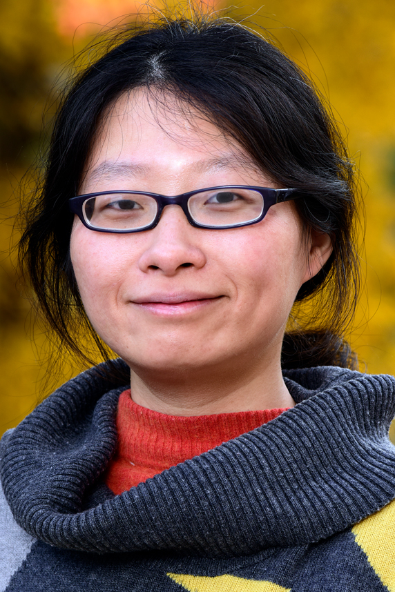
 “Rong Li jumps onto new techniques not just because they’re new but because she has a clear vision of where she wants to go with her science,” DeMayo said of the IRTA Fellow, shown in this picture. “She designs the kitchen — doesn’t just buy the hardware. She knows what she wants to cook.” (Photo courtesy of Steve McCaw / NIEHS)
Fine resolution, finer collaboration
To support embryo development, the uterus undergoes a series of intricate spatial-temporal changes during early pregnancy. Previous studies that used traditional approaches such as histology predicted the existence of certain distinct mouse uterine compartments during early pregnancy. However, due to technical limitations, a systematic analysis of the uterine compartment–specific transcriptome during early pregnancy has never been achieved.
The group used 10x Genomics spatial transcriptomics technology because of its fine resolution at the level of a small group of several cells. The collaborative efforts included sample preparation by Li, sequencing by Xin Xu, Ph.D., from the Epigenomics and DNA Sequencing Core Laboratory, and data analysis and interpretation by Tian-Yuan Wang, Ph.D., from the Integrative Bioinformatics Support Group.
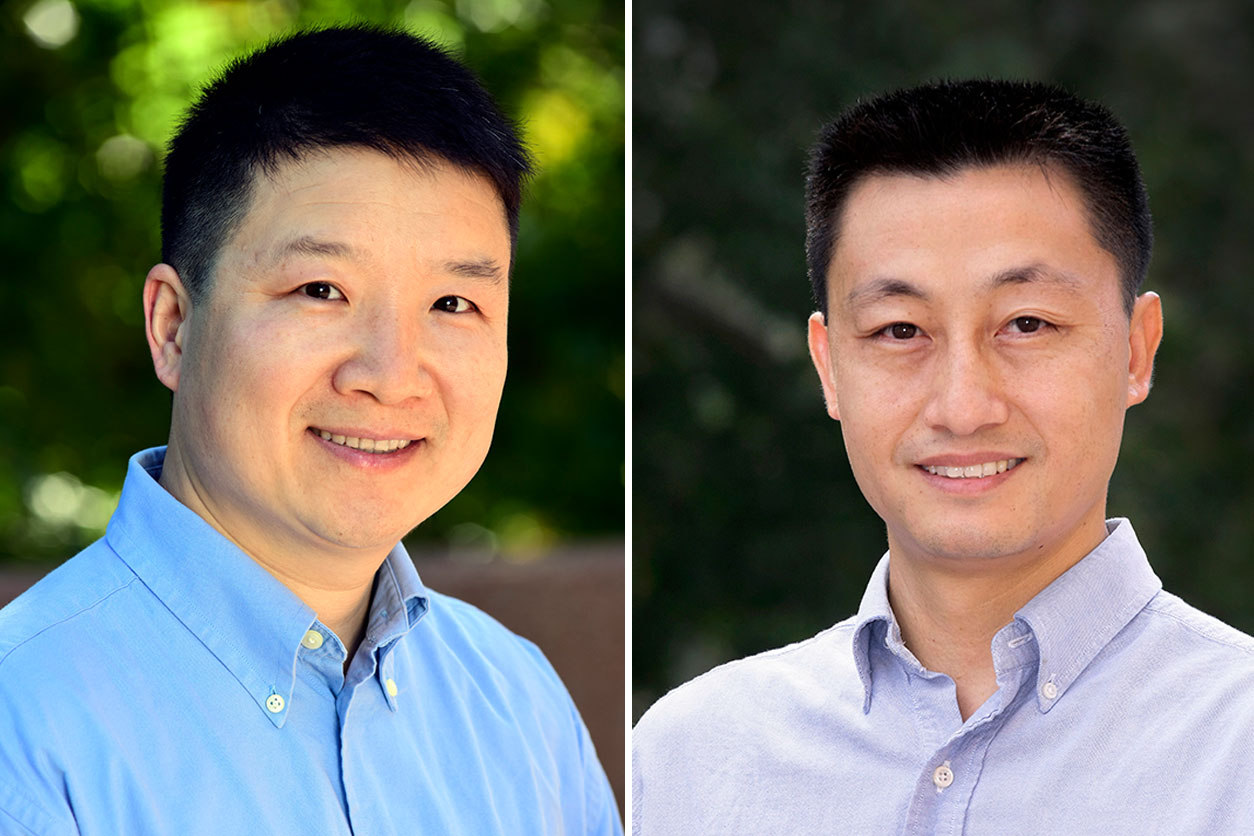
 Xu, left, and Wang, right, played key roles in this research project. (Photo courtesy of Steve McCaw)
“For us, the collaboration of epigenetic core and bioinformatic groups is like giving us a pair of magic glasses that can look through the gross morphology and peep into the delicate molecular world,” Li said.
The study was so effective, others in NIEHS are using the technique.
“Within a year after the successful execution of the first case in March 2021, six groups across NIEHS have utilized or planned to adopt the assay in nine projects,” said NIEHS staff scientist Steve Wu, Ph.D. “Such high demand shows the impact of implementing an assay that facilitates intramural research and ensures NIEHS is at the forefront of scientific endeavors.”
Citation: Li R, Wang TY, Xu X, Emery O, Yi M, Wu SP, DeMayo FJ. 2022. Spatial transcriptomic profiles of mouse uterine microenvironments at pregnancy day 7.5. Biol Reprod; doi:10.1093/biolre/ioac061 [Online 31 March 2022].
(Kelley Christensen is a contract writer and editor for the NIEHS Office of Communications and Public Liaison.)
Source link
factor.niehs.nih.gov

