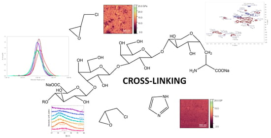
3.1. Cross-Linking
In the experiments in absence of I, where H was cross-linked with E at 1/1, 1/2, and 1/3 molar ratios, the respective obtained yields were 77%, 64%, and 42%. These results indicate that all the H-serine end groups were already fully involved in the reaction at 1/1 of the H/E ratio. This indicates that these conditions were optimal for the 1H/1E ratio to react with the serine-end group to produce the sequence: H-(1→4)-β-D-GlcA-(1→3)-β-D-Gal-(1→3)-β-D-Gal-(1→4)-β-D-Xyl-α-Ser-CH2-CH(OH)-CH2-OH, with the highest observed yield. At the ratio of 1H/2E, we expect that the product with the obtained 64% yield was likely: H-(1→4)-β-D-GlcA-(1→3)-β-D-Gal-(1→3)-β-D-Gal-(1→4)-β-D-Xyl-α-Ser-CH2-CH(OH)-CH2-O-CH2-CH(OH)-CH2-OH. Analogously, the 1H/3E ratio could produce the third 2-hydroxypropyl on the serine-end unit: H-(1→4)-β-D-GlcA-(1→3)-β-D-Gal-(1→3)-β-D-Gal-(1→4)-β-D-Xyl-α-Ser-CH2-CH(OH)-CH2-O-CH2-CH(OH)-CH2-O-CH2-CH(OH)-CH2-OH, which led to the obtained 42% yield.
In the presence of I, when the amount of I was increased at a constant amount of H and E, i.e., at H/E/I ratios of 1/1/1, 1/1/2, and 1/1/3, the obtained yields were 46%, 32%, and 24%. The imidazole content in all products was also confirmed by elemental analysis, indicating an increased nitrogen content (see
Section 2). Since at the H/E/I ratio of 1/1/1, the yield was the highest, we assume that one I molecule is taking part in the reaction with the heparin serine-end unit to form a new sequence (46% yield), as shown in
Figure 1. At the ratio of H/E/I = 1/1/2 (32% yield), the sequence might be formed as shown in
Figure 2.
At the H/E/I ratio of 1/1/3, the remaining imidazolium cation could be attached to some sterically available uronate anion, which led to the lowest yield (24%).
3.2. NMR Analysis
In comparison to
1H NMR spectrum of the unfractionated H, based on our previous studies on H [
4], the 2D NMR analysis method is more informative for the identification of new substituents and it prevents overlapping of the signals. Ten anomeric signals were detected in the HSQC spectrum of H (
Figure 3). This result is also in agreement with the known data [
18]. The signals at 5.42/99.26 ppm (Glc
NS,6S-Ido
2S) and 5.22/102.08 ppm (IdoA
2S-Glc
NS,6S) were the most intense, similar to previous observations and specifications used by Mauri et al. for unfractionated H [
10]. The signals at 5.58/100.30 ppm (Glc
N,6S-GlcA), 5.57/98.92 ppm (Glc
NS,3S,6S), 5.04/104.73 ppm (IdoA
2S-Glc
NS,6S), 4.98/104.57 ppm (IdoA-Glc
NS), 4.66/106.72 ppm (GalA + Gal), 4.54/104.40 ppm (GlcA-Glc
N6,3S,6S), 4.50/105.23 ppm (GlcA-Glc
NAc), and 4.47/105.73 ppm (Xyl-Ser) ppm are minor signals, with the Xyl linked to serine with an α-glycosidic bond at the end of the heparin chain [
10]. The 6 Glc
NS,6S-Ido
2S signal occurs at 4.28, 4.42/68.97 ppm, and the signals of non-sulfated 6 GlcA-Glc
NS,3S,6S are observed at 3.84, 3.88/62.42 ppm [
10]. Additionally, minor CH
2 signals of the 5 Xyl unit occur at 3.41, 4.12/65.83 ppm. The COSY spectrum (not shown here) showed that the signal at 3.92/56.61 ppm belongs to 2 Glc
NAc, the signal at 3.78/85.48 ppm is due to 2 GlcA + Gal, and that of the 3 GlcA + Gal units is at 3.84/84.98 ppm [
10]. The above assignments confirmed the presence of the H-end motif: H-(1→4)-α-D-GlcNS6S-(1→4)-β-D-GlcA-(1→4)-β-D-Gal-(1→3)-β-D-Gal-(1→3)-β-D-Xyl-α-Ser [
10,
21].
The number of anomeric signals is identical for heparin cross-linked with epichlorohydrin at a 1/1 molar ratio at the highest yield (1H/1E; 77% yield) in the HSQC spectrum (
Figure 4) as to that for reference H. The signals at 5.42/100.59 ppm (Glc
NS,6S-Ido
2S) and 5.27/102.06 ppm (Ido
2S-Glc
NS,6S) were the most intense. The presence of the A
NS,3S,6S (5.54/99.20 ppm) and GlcA-Glc
NS,6S signals (4.63/104.52 ppm) indicates that the GlcA units linked to A
NS,6S were not degraded. The 2 Glc
NS,6S-GlcA signal occurs at 3.41/75.67 ppm and 3 Glc
NS,6S-GlcA at 3.40/75.70 ppm. New signals at 3.73, 3.79/48.21 ppm (-
CH2-), 3.46/56.81 ppm, 3.47/59.45 ppm, and 4.06/57.11 ppm [-
CH(OH)], related to the 2-hydroxyprophyl cross-linking bridge (-CH
2-CH(OH)-CH
2-), were observed. The rest of the ring signals were identical to those for the H sample. The hydroxypropyl bridge linking serine, H-(1→4)-β-D-GlcA-(1→3)-β-D-Gal-(1→3)-β-D-Gal-(1→4)-β-D-Xyl-α-Ser-CH
2-CH(OH)-CH
2-OH, is supported by -CH(OH)- signals at 2.92/47.47 ppm, 3.93/56.49 ppm, and 3.46/59.48 ppm in the HSQC spectrum. These three signals might represent central parts of three different mono-functionally linked 2-hydroxipropyl groups. The shape of the bridge is also supported by the fact that there are no interactions between the -CH(OH)- groups at 2.92/47.47, 3.93/56.46, and 3.46/59.48 ppm and any other signal in the corresponding HMBC spectrum of the 1H/1E sample.
When cross-linked at a 1/2 ratio (1H/2E), the yield slightly decreased (64%). The NMR data for 1H/2E were identical to those for 1H/1E. The cross-linking at a 1/3 ratio (1H/3E) decreased the yield even further down to 42%. The HSQC data were also identical to those for 1H/1E.
The HSQC spectrum of the 1H/1E/1I sample (
Figure 5) also revealed ten anomeric signals (pD = 7.40; 46% yield). The second-highest yield was observed for 1H/1E/2I (pD = 5.97; 32%). The anomeric signals were assigned analogously to 1H/1E/1I, i.e., [5.35/100.00 ppm (Glc
NS,6S-IdoA
2S), 5.44/99.60 ppm (GlcA
NAc), 5.24/99.54 ppm (Glc
NAc-GlcA), 5.33/98.38 ppm (Glc
NS-IdoA), 5.57/98.74 ppm (Glc
NS,6S), 5.48/99.40 ppm (Glc
NS-GlcA), 5.56/100.48 ppm (GlcNS,6S-GlcA), 5.14/102.01 ppm (IdoA
2S-Glc
NS,6S), 5.01/104.78 (IdoA-Glc
6S), 4.55/104.19 ppm (IdoA-Glc
NS), 4.67/106.73 ppm (Glc + Gal), 4.61/104.80 ppm (GalA-Glc
NS,6S), 4.62/103.80 ppm (GlcA-Glc
NS), 4.53/105.30 ppm (GlcA-Glc
NAc), and 4.47/105.72 ppm (Xyl-Ser)]. Additionally, two new -CH
2– groups, at 3.66/65.19 and 3.76/63.61 ppm, and four new -CH(OH)- groups, at 2.92/47.42, 3.90/56.39, 3.37/59.54, and 4.59/65.89 ppm, were observed. The first two CH(OH) groups (2.92/47.42 and 3.93/56.49 ppm) were also present in the 1H/1E experiment, while the other two -CH
2– (3.66/65.19 ppm and 3.76/63.61 ppm) and CH(OH) groups observed at 3.37/59.59 ppm and 4.59/65.89 ppm are new, and we assume that they are related to the imidazolium rings.
3.5. AFM and PF-QNM Analysis
Figure 8a–g shows the measured surface morphology of all prepared heparin-based films, represented by 2 × 2 μm
2 scans. Representative corresponding line profiles are also shown. The box and whisker plot in
Figure 8h shows the statistical distribution of the root-mean-square (RMS) surface roughness extracted from several 2 × 2 μm
2 scans acquired at various locations across the surface of the measured films. The evaluation of surface roughness reflects the upper surface of the film after the liquid derivatives were poured into the Petri dishes and dehydrated. Potentially different viscosities of the prepared derivatives may have resulted in slightly different thicknesses of the prepared films; however, we do not expect this to have any significant effect on the resulting determined surface roughness, which was more significantly affected by various particles and pores present on the film surfaces. While no clear trend regarding roughness was observed related to the cross-linking with E and I, the reference heparin showed the widest spread of roughness values, which can be attributed to the large number of pores, ~15–100 nm deep across the surface of the H layer, and also to the presence of various particles (~150–250 nm). Similar pores were only observed in the 1H/3E film; however, they occurred less frequently. Films 1H/2E and 1H/1E/2I showed the lowest surface roughness (~1 nm), which was related to the much-decreased number of spherical particles, which were otherwise present in all the remaining films. The following increasing trend of average surface roughness was observed: 1H/2E (0.9 nm) < 1H/1E/2I (1.0 nm) < 1H/1E (2.1 nm) < 1H/1E/1I (2.2 nm) < 1H/3E (2.3 nm) < H (3.5 nm) < 1H/1E/3I (3.6 nm). This indicates that the most cross-linked films exhibit a decreased roughness of the surfaces when compared to those of H. The best result was observed in the 1H/2E sample, which indicates the optimum result for dimer hydroxypropyl, formed on the serine end-unit. The experiments containing, I exhibit higher roughness values with increasing amounts of I.
The observed surface morphologies of the prepared H-based films showed similarities to those of some of the previously studied seaweed carrageenan films; however, those generally exhibited a higher surface roughness (~2.4–18.6 nm) for scans of similar dimensions [
23]. Some similarities, such as the size of granular features, were also observed in the line profiles. This behavior may be explained by the high density of the strongly charged carboxyl and sulfate groups with relatively low molecular weight (M
w = 11,065 g/mol; M
w/M
n = 1.54) present in the H film in comparison to those of the carrageenans in the films prepared from seaweed extracts. The values at the peak (M
p) were considerably larger for the carrageenans (from 77.8 to 994.0 kg/mol) [
23]. Although the M
w value of the 1H/1E sample increased only slightly (19,900 g/mol; M
w/M
n = 1.54) compared to that for the seaweed polysaccharides, their surface roughness values were significantly different (i.e., higher for the seaweed-based films). The lowest average RMS surface roughness of ~1 nm for the 1H/2E and 1H/1E/2I films was even lower than the results obtained for the seaweed polysaccharides [
14]. The further increase in molecular weight observed for the 1H/1E/1I sample (M
w = 24,200 g/mol; M
w/M
n = 1.56) resulted in a slight increase in the average RMS surface roughness value of ~2.2 nm. All the above surface roughness values are comparable with the values determined for the chitosan/heparin multilayer films [
25], as well as with the RMS values determined on gold covered with PEI cross-linked heparin covered with synthetic polymers used for antidote heparin [
24].
Currently, only a limited number of reports dedicated to the evaluation of the mechanical properties of heparin films [
4,
22,
23], or other polysaccharides [
24,
25,
26], exist. To evaluate the influence of E and I cross-linking on heparin’s mechanical properties, the PeakForce quantitative nanomechanical mapping technique was applied to all prepared films, and the reduced DMT elastic modulus was determined. We note that this technique is limited to probing only the surface or near-surface mechanical properties of the films, and these may differ significantly from those of the bulk, where other mechanical phenomena may play an additional role. Nevertheless, the surface mechanical properties could be important to evaluate, e.g., the wear resistance and longevity of the prepared films in various applications.
The spatially resolved maps of the reduced elastic modulus (
E*) of all the prepared films are shown in
Figure 9a–g. These maps represent the same measurement locations as those shown in the surface morphology scans
Figure 8a–g. The box and whisker plot in
Figure 9h shows a statistical distribution of the
E* values extracted from the scans in
Figure 8a–g, assuming their normal distribution, and are representative for several measurement locations. Here, the green-colored boxes represent the data falling within the lower (Q1) and upper (Q3) quartiles of a probability density function accounting for 50% of all values, i.e., the interquartile range (IQR, Q3-Q1); the mean and median values are also shown. The whiskers represent the minimum and maximum values calculated as Q1 − 1.5 × IQR and Q3 + 1.5 × IQR, respectively. Data falling in the range between the lower and upper whiskers represent 99.3% of all the determined
E* values. The remaining 0.7% of the data points represent outliers with low statistical significance and are depicted with red diamond symbols outside of the whiskers. These cannot be completely avoided during the measurement due to influences such as edge effects near the pores and particles; however, they have a negligible effect on the reported average values and trends.
The
E* maps of investigated films were mostly homogeneous, but showed spatial variations which were typically correlated with the surface morphology, especially near the locations of the pores and particles (
Figure 9). When probing such features, the probe’s tip-to-sample area may vary significantly, which can be reflected in a lower accuracy of the determined values and artifacts, i.e., edge effects, and therefore, such results need to be treated cautiously.
As mentioned previously, one of the significant observed features in almost all samples was the existence of surface particles. Maps of the reduced modulus confirmed that these particles exhibited systematically lower values of ~6–8 GPa for all films, except for the 1H/1E/3I sample, in which their average modulus value was close to that of the whole scanned area. It is therefore likely that the surface particles observed in films H, 1H/1E, 1H/2E, 1H/3E, 1H/1E/1I, and 1H/1E/2I may have a similar origin, and their occurrence is closely related to the cross-linking process used. Also, since the particles were already present in the reference H film, it is likely that their occurrence in the cross-linked films may have originated from heparin, rather than from the cross-linkers used. The
E* map of the 1H/3E film in
Figure 9d showed locations of higher-than-average values which are spatially correlated with the locations of the pores. We believe that rather than functioning as a true material property, these values represent unavoidable artefacts due to the varying contact area inside of the pores and close to their edges.
The following increasing order of the determined mean values of the surface reduced elastic modulus was found: 1H/1E/3I (~7 GPa) < H (~8.2 GPa) <1H/1E/1I = 1H/1E/2I (~8.4 GPa) < 1H/1E (~11.5 GPa) < 1H/2E = 1H/3E (~13.4 GPa). A larger improvement of
E* was found for samples cross-linked with E; however, increased E content did not lead to a considerable increase in
E*. A similar increase was also observed for CCMC in comparison to CMC [
18]. Compared to the reference H cross-linked 1H/1E, the 1H/2E and 1H/3E films showed approximately 40% and 63% higher
E* values. While increasing the E to H ratio to two led to an increase in the reduced modulus from 11.5 GPa to 13.4 GPa, no further significant increase was observed when higher E to H ratios was used, suggesting that the preparation conditions for the 1H/2E film were close to optimal. On the contrary, the films cross-linked with E in the presence of I showed negligible improvement of surface
E* (1H/1E/1I and 1H/1E/2I), or even its decrease by ~15% (1H/1E/3I). This seems to be due to the two limited nitrogen atoms on imidazole available for cross-linking. It is worth mentioning that despite the limited improvement of mechanical properties and the reduced modulus of substances in different ratios, the 1H/1E/1I and 1H/1E/2I films showed the narrowest spread between its values, reflecting improved surface morphology and a low number of particles. The 1H/1E/3I sample, displaying the largest roughness value, exhibits the smallest reduced modulus value.


