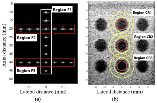
5.1. The Geometric Distortion
The EGSCW, EGSCWe, EGSCLe, and EGSCK equally decrease the average FWHMLat, obtaining a reduction of 0.46 mm (63%), compared to the DAS method, with 0.10 mm (27%) and 0.09 mm (25%) compared to EGSC and GSC techniques, respectively.
The EGSCKe presented the result closest to the actual value, representing a reduction in the average FWHMLat of 0.49 mm (67%), 0.13 mm (35%), and 0.12 mm (33%) compared to DAS, EGSC, and GSC techniques, respectively.
5.2. Contrast
The EGSCWe technique generated a contrast mean value of 52.49 dB, representing an average contrast improvement of 19.66 dB (60%), 19.86 dB (61%), and 22.28 dB (74%) compared to EGSC, GSC, and DAS, respectively. We noticed that the EGSCWe improved the performance of the EGSCW contrast by 1.19 dB, approximately 2.3%.
The EGSCL shows a contrast value close to GSC and EGSC methods and presents an improvement of contrast of 2.35 dB (7.8%) with regard to the DAS technique. However, the EGSCLe decreased the contrast by 3.85 dB (11.7%), 3.65 dB (11.2%), and 1.23 dB (4%) compared to EGSC, GSC, and DAS, respectively.
The EGSCK technique presents the worst contrast value on average, with a reduction of 8.65 dB (26.3%), 8.45 dB (26%), and 6.03 dB (20%) compared to the EGSC, GSC, and DAS, respectively. Also, the EGSCKe shows a slight contrast improvement of 1.1 dB (3.6%) compared to the DAS method.
The EGSCW and EGSCWe methods obtained a reduction in the noise around the fetus, providing better visualization of the contour.
Numerically, the contrast value increased with these techniques compared to the others. These methods showed improvement of 3.59 dB (9%) and 4.02 dB (10%), respectively, compared to the DAS method. An improvement, in contrast, allows better visualization of different areas, densities, and contours, being a positive result.
The comparison between the simulation and in situ experimental results reveals strong consistency, in terms of contrast. While simulations provide valuable theoretical data, the in situ experiments confirmed the effectiveness of techniques such as the EGSCWe and EGSCW, which improved image quality in both simulations and practical conditions. The ability to achieve similar improvements in both domains suggests that these methods are robust, highlighting the potential applicability of these methods in real clinical scenarios.
The geometric distortion results with the EGSCL and EGSCLe methods presented an improvement in the FWHM for the simulation and phantom, compared to EBGSC, GSC, and DAS. In the contrast results, for simulation and phantom images, the regions closest to the transducer suffer a slight homogenization due to filtering, which decreases contrast, especially for the EGSCLe. However, EGSCL and EGSCLe generally presented values around the MVGSC and EGSC methods, indicating future research with these filters.
The EGSCK and EGSCKe methods showed excellent FWHM reductions for simulation and phantom data compared to the DAS, MVGSC, EGSC, EGSCL, and EGSCLe methods. However, in terms of contrast, the EGSCKe method presented values close to the DAS, and the EGSCK presented a significant decrease with the lowest contrast values, which may indicate that there are better techniques to improve this parameter.
Compared to conventional methods, the improvement in contrast and reduction in distortion could represent better visualization of tissues, detection of small targets, and greater detail of the areas analyzed, which is positive for clinical use, helping with the quality of B-mode images and also in imaging modes that require high framerate, such as elastography, ultrafast Doppler, and ultrasound localization microscopy (ULM).
The sizes of the signal subspace functions in the beamformer eigenspace and the windows for the adaptive filters can present different contrast and lateral distortion results, which can help analyze different regions and need to be better studied.
In this work, we did not do tests with 3D Ultrafast images. However, as the filtering techniques improved the resolution, they can be applied to each processed frame before reconstructing the high-resolution 3D images.
Source link
Larissa C. Neves www.mdpi.com

