3.1. Enzyme Activity Profiles
The identities of
T. reesei RUT C-30 and
A. brasiliensis were confirmed at the molecular level, with representative sequences deposited in NCBI as described in
Section 2. In addition to investigating enzymatic induction during co-culture over time, this study also examined monocultures and the impact of different colonization time periods between both fungi in co-cultivation, as summarized in
Table 1.
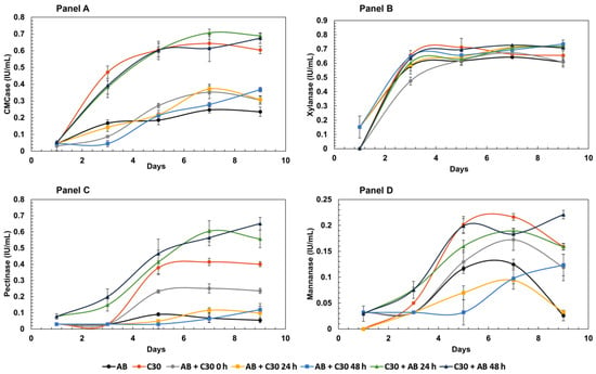
Figure 1 displays enzymatic activities using xylan, mannan, carboxymethylcellulose (CMC) and pectin. The highest CMCase activity was observed for co-cultures of
T. reesei RUT-C30 where
A. brasiliensis inoculation was staggered for 24 h and 48 h (C30 + AB 24 h and C30 + AB 48 h, respectively). Considering the standard deviations, it can be concluded that both co-cultures produced similar results. Performances for both co-culture models were approximately 10% higher than observed for the
T. reesei RUT-C30 monoculture at the final nine-day time point. One possible explanation for such findings is that β-glucosidase from
A. brasiliensis may supplement the typical low production of this enzyme by
T. reesei RUT-C30. Such a supplementation might prevent events such as CMCase inhibition by its hydrolysis products, thereby enhancing the release of reducing sugars.
Previous studies have shown an increase in cellulase activities in co-cultures of
T. reesei RUT-C30 with other Aspergillus fungi, including
A. niger, under different cultivation conditions [
17]. Zhao et al. [
18] also demonstrated a 40% increase in CMCase activity in co-cultures of
T. reesei RUT-C30 and
A. niger compared to the monoculture of
T. reesei. Regarding xylanase activity, minimal differences were observed among the tested cultures, and from the fourth day onwards, activities of all cultures became indistinguishable. On the other hand, pectinase activity of co-cultures of
T. reesei RUT-C30 inoculated with
A. brasiliensis after 24 h and 48 h exhibited a notable performance, similar to that observed for CMCase activity. These co-cultures displayed pectinase activities that were 37% and 48% higher than the
T. reesei RUT-C30 monoculture, respectively. Considering the nearly absent production of pectinases by the monoculture of
A. brasiliensis, these co-cultures demonstrated a 700% increase in this activity when compared to the
A. brasiliensis monoculture. Such activities described here are relevant, given that pectinases are enzymes with considerable importance for the fruit pulp and juice industry [
19], as well as with applications in tea, coffee and even textile industries [
20]. Mannanase activity showed the lowest overall performance among the mono- and co-cultures. Co-culture of
T. reesei RUT-C30 inoculated after 48 h with
A. brasiliensis exhibited noteworthy results, however, with values close to 0.25 IU/mL. The lower values for pectinase and mannanase activities can be attributed to the composition of sugarcane bagasse, which contains smaller amounts of their respective substrates within the sugarcane bagasse cell wall.
When evaluating activities tested on synthetic substrates, the co-cultures demonstrated significant β-glucosidase, β-xylosidase, and β-galactosidase activities (
Figure 2). As expected, the monoculture of
T. reesei RUT-C30 displayed the lowest β-glucosidase activity, a well-known characteristic for this strain that is widely reported in the literature [
3,
21]. Conversely, the monoculture of
A. brasiliensis exhibited a 57% higher β-glucosidase activity than
T. reesei RUT-C30 on the final day of analysis.
A. brasiliensis, given its ability to produce this enzyme, offers potential as a non-mycotoxin-producing alternative to the commonly employed
A. niger, which is often partnered with
T. reesei RUT-C30 to enhance β-glucosidase activity in co-cultivation cocktails [
18,
22]. The co-culture format that exhibited the greatest potential for increasing β-glucosidase activity was achieved through simultaneous inoculation of the two fungi (AB + C30 0 h), resulting in an intermediate level between the activities of
A. brasiliensis and
T. reesei RUT-C30, with a 38% increase compared to the latter fungus in monoculture. When comparing inoculation delay experiments on β-glucosidase activity, where
T. reesei RUT-C30 was the first colonizer, a negative impact on enzyme activity was observed. Experiments with
A. brasiliensis inoculated with delays of 24 h and 48 h compared to
T. reesei RUT-C30 showed decreases in β-glucosidase activity on the final day of cultivation. For the co-culture where
T. reesei RUT-C30 was employed as the first colonizer, followed by
A. brasiliensis after 24 h (C30 + AB 24 h), a significant increase in enzymatic activity was observed between days 5 and 7, although this value dropped substantially on the final day of incubation.
A. brasiliensis monoculture resulted in the highest β-xylosidase activity, consistent with previous studies that have demonstrated its ability to produce this enzyme when grown on lignocellulosic substrates [
23]. Co-cultures of
A. brasiliensis, with
T. reesei RUT-C30 inoculated after 48 h, resulted in β-xylosidase activity values similar to those of the monoculture of
A. brasiliensis. The co-culture of simultaneous inoculation (AB + C30 0 h) performed similarly to that of the monoculture of
T. reesei RUT-C30, secreting approximately 0.25 IU/mL. Co-culture with a 48 h delay resulted in β-xylosidase activities 696% higher than observed in simultaneous cultivation and monoculture of
T. reesei RUT C-30. One possible explanation for this increase in activity may reflect the close evolutionary relationship between
A. brasiliensis and
A. niger. The latter species is known to acidify growth media to create preferred growing conditions. This is particularly common in the absence of competition and during the initial stages of cultivation, with reports of acidification also influencing enzymatic activities [
24].
A. brasiliensis may potentially share a similar capacity. These results also highlight the impact of the time intervals between inoculations. Cultures of
A. brasiliensis subsequently inoculated with
T. reesei RUT-C30 after a shorter interval (24 h) exhibited a completely different β-xylosidase induction curve, with a sharp drop in activity on the ninth day of cultivation. These data suggest influencing effects on the first day when
T. reesei RUT-C30 is present, decreasing β-xylosidase activity. The negative effect of
T. reesei RUT-C30 on β-xylosidase activity is further confirmed by the other experimental setup where
T. reesei RUT-C30 is the initial colonizer, with
A. brasiliensis subsequently inoculated. In all cultures where
T. reesei RUT-C30 is cultivated for a longer period than
A. brasiliensis, β-xylosidase activities were lower than when the opposite scenario was employed. Unfortunately, the available literature on β-xylosidase activities in fungal co-cultures is limited. One of the few investigations of β-xylosidases in co-cultures is the study by Hu et al. [
25], where the co-culture between
A. niger and
A. oryzae resulted in a 55% decrease in β-xylosidase activity when compared to the monoculture of
A. niger. The scarcity of information regarding this enzymatic activity in co-cultures not only hinders direct comparisons but also underscores the significance of the present study for co-cultures.
Regarding β-galactosidase, the AB + C30 48 h co-culture showed the highest activity. Similar to β-glucosidase activities and, to a lesser extent, β-xylosidase, there was also a peak in activity on the seventh day of cultivation. Due to logistical reasons, all the characterizations mentioned in the following sections of this study were conducted using samples from the final day, even though it may not always be the day with the highest activity, as observed for β-glucosidase. Finally, no significant activity for β-mannosidases was detected, similar to the findings in the monoculture experiments for A. brasiliensis.
3.2. Effect of Temperature on the Enzymatic Activity
Crude extract derived from co-cultures is a complex mixture that represents the contributions from each of the different fungi present. Currently, there is limited information in the literature regarding how physical or chemical conditions can affect the enzymatic activity of co-cultures.
The catalytic activity of enzymes can be affected by the environmental temperature. When considering crude fungal extracts, enzyme activities are measured for not just one enzyme but rather a set of enzymes that are acting on the substrate. Regarding co-cultures, this characteristic is further amplified since the crude extract consists of a combination of enzymes produced by the two participating fungi. As such, when evaluating the effect of temperature, what is actually measured is how this physical condition interferes globally with the activity of a specific group of enzymes. Investigating such parameters is important from an industrial standpoint, since commercially available enzyme cocktails are typically composed of crude extracts with the possible addition of exogenous enzymes, rather than purified enzymes alone.
Figure 3 depicts the effect of temperature on the enzymatic activity of the co-cultures. Temperatures ranging from 30 to 80 °C were tested to encompass conditions relevant to industrial applications.
Regarding CMCase activity, the monoculture of T. reesei RUT-C30 maintained the highest activity between 30 and 50 °C, with an activity peak observed at 50 °C. On the other hand, the CMCase activity of C30 0 h was surpassed by AB + C30 0 h at 60 °C and by AB monoculture at 70 °C and 80 °C. As far as we are aware, this is the first report on the effect of temperature on CMCase activity in A. brasiliensis.
Xylanase activity showed the lowest variation according to temperature across all examined cultures. This result was expected, considering that several xylanases which are active at high temperatures have already been reported in the literature, most of which exhibit an optimal activity in the range of 40–60 °C [
26]. Concerning co-cultures, AB + C30 0 h produced high xylanase activities between 30 °C and 60 °C which are only comparable to the C30 monoculture.
Regarding pectinase activity, the highest values were obtained by C30 monoculture and AB + C30 0 h, both at 70 °C. T. reesei RUT-C30 also presented the highest values for mannanases when assays were conducted at 70 °C. Interestingly, C30 + AB 24 h and C30 + AB 48 h also showed activity comparable to C30 at 70 °C and higher at 80 °C. Overall, mannanase activities obtained in mono- and co-culture formulations were low, indicating that sugarcane bagasse is not, at least under the conditions presented here, an efficient inducer of mannanase activity for the characterized fungi.
The practice of employing a fungus capable of producing β-glucosidase as a partner to supplement
T. reesei RUT-C30’s deficiency in production of this enzyme is a co-cultivation practice that has been employed with varying degrees of success [
18,
27,
28]. Here,
A. brasiliensis monoculture showed the highest β-glucosidase activity at a temperature of 50 °C. It is interesting to note that β-glycolytic activity in
A. brasiliensis remained constant at 50 and 60 °C, with activity in co-culture (AB + C30 0 h) approximately 30% higher at 60 °C than at 50 °C. This fact, along with several other situations presented here, reinforces the conclusion that fungal co-cultures can show unique, complex, and, so far, unpredictable enzymatic characteristics, which are not merely averages between the activities of each of the participating fungi but rather a new composition of the enzymatic cocktail. Interestingly, all cultures where
T. reesei RUT-C30 was inoculated at intervals of 24 and 48 h prior to
A. brasiliensis resulted in low β-glucosidase enzyme activities.
β-xylosidase activity was the highlight for
A. brasiliensis in terms of enzyme secretion. In monoculture, values reached approximately 6 IU/mL at 70 °C, the highest enzyme activity compared to C30 monoculture and all co-culture configurations. In support of our findings, as mentioned earlier, the presence of thermophilic β-xylosidases has also been observed in
A. brasiliensis grown on agro-industrial residues [
29].
For β-galactosidase activity, the highest activities were observed in the co-culture AB + C30 48 h throughout the temperature range. Therefore, a clear synergistic effect of the interaction between both fungi could be observed, with the activity achieved by the co-culture higher than the sum of the activities of the monocultures. With greatest activity at 60 °C, the AB + C30 48 h co-culture showed an activity 46% higher than the sum of the individual activities of
A. brasiliensis and
T. reesei RUT-C30 monocultures. The same synergistic effect was also evident, to a lesser extent, at temperatures of 30, 40, and 50 °C. From an industrial point of view, the presence of β-galactosidases that are active at 30 °C or lower temperatures is interesting, as these are typical conditions applied in the dairy industry, where these enzymes are widely employed. The AB + C30 48 h co-culture can be seen as a potential new source of β-galactosidases, enzymes which have enormous economic potential in the food industry [
30].
3.3. Effect of pH on Enzymatic Activity
Given its potential importance in co-culture, the effect of pH on the activity of CMCase in the mono- and co-cultures was also investigated, with a pH range between 3 and 10 examined, across one-unit intervals (
Figure 4). Once pH conditions were controlled, the simultaneous co-cultivation of
A. brasiliensis and
T. reesei RUT-C30 (AB + C30 0 h) showed the highest total values among the cultures. The AB + C30 0 h co-cultivation performed better in the more acidic pH range, as expected and already well documented for enzymes from filamentous fungi. The highest activity of this culture was achieved at pH 4, where a CMCase activity of 0.74 UI/mL was observed. This activity was 32% higher than observed in the monoculture of
T. reesei RUT-C30, a fungus renowned for its CMCase activity. Throughout the acidic pH range, i.e., pH 3, 4, 5, and 6, the AB + C30 0 h co-culture obtained activities 25%, 32%, 25%, and 33% higher than observed for the monoculture of
T. reesei RUT-C30, respectively.
T. reesei RUT-C30 is a strain known for its low production of β-glucosidase, while
A. brasiliensis has shown to be a potential producer of this enzyme. In this way, the simultaneous cultivation of these two fungi may have created a situation of enzymatic complementarity, resulting in the possible synergy that increased hydrolytic efficiency. Synergy between the various cellulase activities such as endocellulases (CMCases), cellobiohydrolases, and β-glucosidases is crucial for the deconstruction of the cellulose polymer [
18]. Therefore, it is possible that the β-glucosidase from
A. brasiliensis alleviated the product inhibition caused by cellobiose in other cellulolytic enzymes, thus increasing the efficiency of CMC degradation in this experiment.
3.5. Proteomic Analysis of Mono- and Co-Cultures
As summarized above, specific enzyme assays enabled a comparison of biomass-degrading enzyme activities in A. brasiliensis and T. reesei RUT-C30 mono- and co-cultures. However, this hypothesis-driven approach certainly misses important global information on the protein composition of the samples. Proteomics, a high-throughput and usually a discovery-driven strategy, can provide a qualitative and quantitative description of the secretomes (i.e., the set of proteins expressed by an organism and secreted into the extracellular space) of the mono- and co-cultures studied here.
Thus, we performed a label-free quantitative bottom-up secretome analysis of the final (9 days) CE of each sample for the samples AB, C30, AB + C30 0 h, AB + C30 24 h and C30 + AB 24 h (see
Table 1).
For all samples, including those originating from monocultures of
A. brasiliensis and
T. reesei RUT-C30, protein identification searches were based on a concatenated database composed of both
T. reesei RUT-C30 and
A. brasiliensis reference proteomes along with common contaminants. As mentioned earlier, this strategy revealed a total of 167 proteins, with 78 from
T. reesei RUT-C30 and 89 from
A. brasiliensis. (
Supplementary Table S1).
The specificity of the identification strategy was validated by evaluating the list of proteins identified from C30 and AB monocultures. Remarkably, for both monoculture secretomes, all identified proteins (below 1% FDR cutoff) corresponded to UniProt accessions belonging to their specific organism (
Figure 5). The absence of cross-species protein identification demonstrates the specificity of the data generated for mono- and co-culture secretomes.
In the AB + C30 0 h co-culture, approximately 75% of the identified proteins belonged to
T. reesei RUT-C30, while
A. brasiliensis accounted for around 25% of the identified proteins. This suggests a greater diversity and/or a higher abundance of secreted
T. reesei RUT-C30 proteins compared to
A. brasiliensis. By contrast, when
T. reesei RUT-C30 was inoculated 24 h after
A. brasiliensis (AB + C30 24 h), the proportion of identified proteins from each organism was approximately 50%, indicating a similar diversity and more comparable abundance of proteins between the two organisms (
Figure 5). Interestingly, when
A. brasiliensis was inoculated 24 h after
T. reesei RUT-C30 (C30 + AB 24 h), proteins from
A. brasiliensis were not identified. This could be explained by a higher growth rate of
T. reesei RUT-C30 compared to
A. brasiliensis. However, we observed in monocultures that the growth rate of both fungi is similar. It cannot be ruled out that
T. reesei RUT-C30, which is a genetically improved organism, may display a better adaptation to the competitive conditions of co-cultures.
Differences in protein composition between the co-cultures AB + C30 0 h, AB + C30 24 h and C30 + AB 24 h versus single cultures were represented using Venn diagrams (
Figure 6). Additionally, an inset table shows the proteins specifically found in the co-cultures (six for AB + C30 0 h, seven for AB + C30 24 h and five for C30 + AB 24 h).
Figure 6C shows that C30 + AB 24 h displays a high number of proteins (34) in common with C30 but with no overlap with AB. This pattern is different to that observed in comparisons with AB + C30 0 h (
Figure 6A) and AB + C30 24 h (
Figure 6B), which show proteins in common with AB (10 and 27, respectively), although a higher overlap was observed against C30 (30 and 26, respectively).
The quantification of the 167 proteins was evaluated by extracted ion chromatograms (XIC) from identified peptides. These were further annotated with match between runs to minimize the number of missing values in each run. This approach relies on the propagation of identification from peptides with similar characteristics in different runs and is highly appreciated for quantitative proteomic comparisons. However, it is important to note that some identification propagation may be misleading, especially in the absence of MS/MS information. To mitigate potential effects, statistical tests were performed only between conditions with similar protein/peptide matrices, namely AB + C30 0 h and AB + C30 24 h. Given the substantial differences in proteins identified when comparing mono- and co-cultures, and particularly in the co-culture condition where
T. reesei was inoculated first (C30 + AB 24 h), resulting in the identification of only
T. reesei proteins (
Figure 5), these datasets were not submitted to ANOVA tests. Nevertheless, global analysis, such as the probabilistic principal component analysis (PPCA), was employed to assess the similarities among replicates within each group and to identify differences between conditions (
Figure 7). PPCA revealed a distinct grouping of all condition replicates, effectively separating single cultures from co-cultures, except for the
T. reesei RUT-C30 single culture (C30) and C30 + AB 24 h co-culture. The similarity between both samples corroborates the substantial overlap in the identified proteins between the C30 + AB 24 h co-culture and C30 monoculture (
Figure 6B). In addition, it was shown that all proteins identified in C30 + AB 24 h belonged to
T. reesei RUT-C30 (
Figure 5). Overall, the PPCA demonstrated differences between conditions and suggested similarities between C30 and C30 + AB 24 h conditions.
Quantitative comparisons were conducted to assess the differences between different time points of
T. reesei RUT-C30 inoculum in the co-culture (AB + C30 0 h vs. AB + C30 24 h). The results of this test revealed 58 regulated proteins (ANOVA,
p-value < 0.05), with 23 proteins up-regulated in AB + C30 0 h and 35 proteins up-regulated in AB + C30 24 h (
Supplementary Table S2). The proteins that showed significantly higher abundance in the AB + C30 0 h condition belonged exclusively to
T. reesei RUT-C30. In contrast, the up-regulated proteins in AB + C30 24 h originated from
A. brasiliensis. These findings are consistent with the identification evaluations, which suggested a higher ratio of
T. reesei RUT-C30-identified proteins in the co-culture with simultaneous inoculation (AB + C30 0 h), as shown in
Figure 5.
Collectively, these data indicate that, not only in terms of diversity but also in terms of abundance,
T. reesei RUT-C30 proteins prevail over
A. brasiliensis when the inoculum occurs simultaneously. On the other hand, when
T. reesei RUT-C30 inoculum is introduced 24 h later,
A. brasiliensis may contribute in a greater proportion to the co-culture, both in terms of diversity and abundance of its secreted proteins. Furthermore, analysis of interactions and functional characterization of the regulated proteins revealed that they were significantly involved in polysaccharide metabolic and catabolic processes (see
Supplementary Table S3). This is evident from the interactions formed among these regulated proteins, including glucanases, mannanases, and beta-glucosidases from
A. brasiliensis in the AB + C30 24 h condition, and alpha-galactosidases, xyloglucanases, endopolygalacturonases, among others, from
T. reesei RUT-C30 in the AB + C30 0 h secretome. Additionally, two regulated proteins in
A. brasiliensis were found to be related to serine proteases (see
Supplementary Table S3). These classifications suggest that
T. reesei RUT-C30 may contribute less to the polysaccharide metabolic processes when this fungus is inoculated 24 h after
A. brasiliensis. However, this decrease in the contribution of
T. reesei RUT-C30 might be compensated for and even enhanced by polysaccharide metabolic enzymes from
A. brasiliensis.


