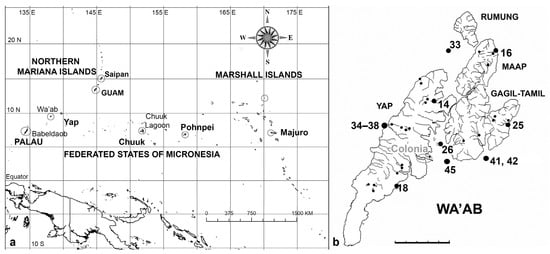
3. Results
Checklist of Yap records to date from this paper and [25] in alphabetical order, listing new records (Y = yes) for Yap and Micronesia in this paper, new species and nomenclatural changes in boldface. The second column indexes the subsection number of the species in the systematic order presented below.
Checklist of Yap records to date from this paper and [25] in alphabetical order, listing new records (Y = yes) for Yap and Micronesia in this paper, new species and nomenclatural changes in boldface. The second column indexes the subsection number of the species in the systematic order presented below.
| Taxon | Subsection Number | New Record for Yap | New Record for Micronesia |
|---|---|---|---|
| Achnanthes armillaris | 94 | Y | |
| Achnanthes cf. brevipes | 95 | Y | |
| Achnanthes grunowii | 97 | Y | Y |
| Achnanthes kuwaitensis | 96 | Y | Y |
| Achnanthes orientalis | 98 | Y | Y |
| Achnanthes parvula | 99 | Y | |
| Actinocyclus decussatus | 5 | Y | |
| Actinocyclus subtilis | 6 | Y | |
| Amphora arenaria | 145 | Y | |
| Amphora bigibba | 146 | Y | |
| Amphora bigibba var. interrupta | 147 | Y | Y |
| Amphora hyalina | 148 | ||
| Amphora immarginata | 149 | Y | |
| Amphora obtusa | 150 | Y | |
| Amphora ostrearia var. vitrea | 151 | Y | |
| Amphora cf. proteus | 152 | Y | |
| Amphora spectabilis | 153 | Y | |
| Amphora subhyalina | 154 | Y | |
| Anaulus minutus | 12 | Y | Y |
| Anorthoneis [eurystoma] sp. | 101 | Y | |
| Ardissonea densistriata | 21 | ||
| Ardissonea formosa | 22 | ||
| Ardissoneopsis appressata | 23 | ||
| Ardissoneopsis gracilis | 24 | ||
| Asterolampra marylandica | 8 | Y | |
| Auricula complexa | 186 | ||
| Auricula flabelliformis | 187 | Y | |
| Auricula intermedia | 188 | Y | |
| Bacillaria “paradoxa” | 159 | Y | |
| Bacteriastrum furcatum | 13 | Y | |
| Berkeleya rutilans | 106 | Y | |
| Biddulphia biddulphiana | 32 | Y | |
| Biddulphiella cuniculopsis | 33 | Y | Y |
| Biddulphiella tridens | 34 | Y | |
| Biddulphiopsis membranacea | 35 | Y | |
| Bleakeleya notata | 44 | Y | |
| Caloneis egena | 115 | Y | |
| Caloneis ophiocephala | 116 | Y | Y |
| Caloneis cf. petitiana | 117 | Y | Y |
| Campylodiscus brightwellii | 189 | ||
| Campylodiscus giffenii | 190 | Y | |
| Campylodiscus humilis | 191 | Y | |
| Campylodiscus imperialis | 192 | Y | |
| Campylodiscus neofastuosus | 193 | ||
| Campylodiscus ralfsii | 194 | ||
| Campylodiscus tatreauae | 195 | Y | Y |
| Campylodiscus wallichianus | 196 | ||
| Chaetoceros peruvianus | 14 | Y | |
| Climaconeis lorenzii | 107 | ||
| Climaconeis minaegensis | 108 | ||
| Climaconeis tarangensis | 109 | ||
| Climacosphenia elegantissima | 25 | Y | |
| Climacosphenia scimiter | 26 | Y | |
| Cocconeis convexa | 102 | Y | |
| Cocconeis coronatoides | 103 | Y | |
| Cocconeis dirupta | 104 | Y | |
| Cocconeis heteroidea | 105 | Y | |
| Colliculoamphora gabgabensis | 82 | Y | |
| Coronia ambigua | 197 | Y | |
| Coronia decora | 198 | Y | |
| Coronia decora var. pinnata | 199 | Y | |
| Cyclophora minor | 64 | Y | |
| Cyclophora tenuis | 65 | Y | |
| Cymatoneis belauensis | 134 | Y | Y |
| Cymatoneis sulcata | 133 | Y | |
| Cymatoneis yapensis | 135 | Y | Y |
| Cymatonitzschia marina | 160 | Y | Y |
| Dictyoneis marginata | 92 | Y | Y |
| Diploneis carolinensis | 118 | ||
| Diploneis cerebrum | 119 | Y | |
| Diploneis chersonensis | 120 | Y | |
| Diploneis claustra | 121 | Y | |
| Diploneis crabro | 122 | Y | |
| Diploneis crabro var. excavata | 123 | Y | |
| Diploneis craticula | 124 | Y | |
| Diploneis denticulata | 125 | Y | Y |
| Diploneis nitescens | 126 | Y | |
| Diploneis papula | 127 | Y | |
| Diploneis smithii | 128 | Y | |
| Diploneis smithii var. rhombica | 129 | Y | |
| Diploneis suborbicularis | 130 | Y | |
| Diploneis weissflogii | 131 | Y | |
| Diploneis weissflogiopsis | 132 | Y | |
| Disymmetria excentrica | 15 | ||
| Disymmetria reticulata | 16 | ||
| Divergita biformis | 46 | Y | |
| Ehrenbergiopsis hauckii | 4 | Y | |
| Entomoneis yudinii | 182 | Y | Y |
| Epithemia guettingeri | 183 | Y | |
| Epithemia muscula | 184 | Y | |
| Falcula paracelsiana | 47 | Y | |
| Gato hyalinus | 80 | Y | |
| Glyphodesmis acus | 81 | Y | Y |
| Gomphonemopsis littoralis | 93 | Y | |
| Gomphotheca marciae | 161 | ||
| Grammatophora angulosa | 58 | Y | |
| Grammatophora oceanica | 59 | Y | |
| Grunowago pacifica | 27 | Y | |
| Halamphora exigua | 155 | ||
| Halamphora turgida | 156 | ||
| Hemiaulus chinensis | 11 | Y | Y |
| Hendeyella lineata | 48 | Y | |
| Hendeyella rhombica | 49 | Y | Y |
| Homoeocladia celaenopsis | 162 | Y | |
| Homoeocladia coacervata | 166 | Y | |
| Homoeocladia martiana | 168 | Y | |
| Homoeocladia micronesica | 167 | ||
| Homoeocladia radiata | 163 | ||
| Homoeocladia schefteropsis | 165 | ||
| Homoeocladia tarangensis | 169 | ||
| Homoeocladia vittaelatae | 164 | ||
| Homoeocladia volvendirostrata | 170 | Y | |
| Hyalosira pacifica | 60 | ||
| Hyalosira tropicalis | 61 | Y | |
| Hyalosynedra laevigata | 50 | Y | |
| Hydrosilicon mitra | 200 | Y | |
| Lampriscus shadboltianus | 36 | Y | |
| Licmophora flabellata | 66 | Y | |
| Licmophora cf. hastata | 67 | Y | Y |
| Licmophora johnwestii | 68 | ||
| Licmophora peragallioides | 69 | Y | |
| Licmophora remulus | 70 | Y | |
| Licmophora romuli | 71 | Y | |
| Licmophora undulata | 72 | Y | |
| Lioloma delicatulum | 75 | Y | Y |
| Lioloma elongatum | 76 | Y | Y |
| Lyrella clavata | 83 | Y | |
| Lyrella lyra | 84 | Y | |
| Lyrella cf. rudiformis | 85 | Y | Y |
| Mastogloia acutiuscula var. elliptica | |||
| Mastogloia affirmata | |||
| Mastogloia amoyensis | |||
| Mastogloia angulata | |||
| Mastogloia binotata | |||
| Mastogloia citrus | |||
| Mastogloia cocconeiformis | |||
| Mastogloia corsicana | |||
| Mastogloia cribrosa | |||
| Mastogloia crucicula | |||
| Mastogloia crucicula var. alternans | |||
| Mastogloia cuneata | |||
| Mastogloia cf. cyclops | |||
| Mastogloia davisii | |||
| Mastogloia delicatissima | |||
| Mastogloia emarginata | |||
| Mastogloia erythraea | |||
| Mastogloia fimbriata | |||
| Mastogloia graciloides | |||
| Mastogloia horvathiana | |||
| Mastogloia hustedtii | |||
| Mastogloia inaequalis | |||
| Mastogloia kjellmanii | |||
| Mastogloia lunula | |||
| Mastogloia mauritiana | |||
| Mastogloia mediterranea | |||
| Mastogloia neoborneensis | |||
| Mastogloia ovata | |||
| Mastogloia ovulum | |||
| Mastogloia paradoxa | |||
| Mastogloia peracuta | |||
| Mastogloia pulchella | |||
| Mastogloia punctatissima | |||
| Mastogloia pusilla var. subcapitata | |||
| Mastogloia quinquecostata | |||
| Mastogloia rhombica | |||
| Mastogloia sergiana | |||
| Mastogloia singaporensis | |||
| Mastogloia sulcata | |||
| Mastogloia tridacnula | |||
| Mastogloia umbra | |||
| Mastogloia varians | |||
| Mastogloia witkowskii | |||
| Mastogloiopsis biseriata | 89 | Y | |
| Microtabella interrupta | 62 | Y | |
| Microtabella rhombica | 63 | Y | |
| Moreneis cf. hexagona | 86 | Y | |
| Navicula consors | 136 | Y | |
| Navicula plicatula | 137 | Y | |
| Navicula tsukamotoi | 138 | Y | |
| Neofragilaria anomala | 38 | Y | |
| Neosynedra provincialis | 53 | Y | |
| Neosynedra tortosa | 54 | Y | |
| Nitzschia frustulum | 171 | Y | |
| Nitzschia longissima | 172 | Y | |
| Nitzschia maiae | 173 | Y | |
| Nitzschia marginulata var. didyma | 174 | Y | |
| Nitzschia obtusa var parva | 175 | Y | Y |
| Nitzschia pseudohybridopsis | 176 | Y | Y |
| Nitzschia ventricosa | 177 | Y | |
| Odontella obtusa | 19 | Y | Y |
| Opephora pacifica | 57 | Y | Y |
| Paralia longispina | 3 | Y | |
| Parlibellus biblos | 110 | Y | |
| Parlibellus hamulifer | 111 | Y | |
| Parlibellus waabensis | 112 | Y | |
| Perideraion montgomeryi | 45 | Y | |
| Perissonoë crucifera | 42 | Y | |
| Petrodictyon gemma | 201 | Y | Y |
| Petrodictyon patrimonii | 202 | Y | |
| Petroneis granulata | 87 | Y | |
| Petroneis humerosa | 88 | Y | |
| Plagiogramma porcipellis | 39 | Y | |
| Plagiogramma subatomus | 40 | ||
| Plagiotropis lepidoptera | 141 | Y | |
| Planothidium delicatulum | 100 | Y | Y |
| Pleurosigma simulacrum | 142 | Y | |
| Podocystis adriatica | 73 | Y | |
| Podocystis spathulata | 74 | Y | |
| Podosira hormoides | 1 | Y | |
| Podosira montagnei | 2 | Y | |
| Proboscia alata | 9 | Y | |
| Progonoia diatreta | 113 | Y | |
| Progonoia intercedens | 114 | Y | |
| Protokeelia cholnokyi | 185 | Y | |
| Psammodictyon constrictum | 178 | Y | |
| Psammodictyon panduriforme | 179 | Y | |
| Psammodictyon pustulatum | 180 | Y | |
| Psammodiscus nitidus | 41 | Y | |
| Pseudictyota dubia | 20 | Y | |
| Rhaphoneis castracanei | 43 | Y | |
| Rhizosolenia imbricata | 10 | Y | Y |
| Rhoicosigma parvum | 143 | Y | |
| Roperia cf. tesselata | 7 | Y | |
| Schizostauron cf. trachyderma | 144 | Y | |
| Skeletonema grevillei | 17 | Y | Y |
| Striatella unipunctata | 37 | Y | |
| Stricosus cardinalii | 51 | Y | |
| Stricosus harrisonii | 52 | Y | |
| Synedra lata | 55 | Y | |
| Synedrosphenia gomphonema | 28 | ||
| Synedrosphenia licmophoropsis | 29 | ||
| Tabularia parva | 56 | Y | |
| Tetramphora decussata | 90 | Y | |
| Tetramphora intermedia | 91 | Y | |
| Thalassionema baculum | 77 | ||
| Thalassionema synedriforme | 78 | Y | Y |
| Thalassiophysa hyalina | 157 | Y | |
| Thalassiothrix gibberula | 79 | Y | Y |
| Toxarium hennedyanum | 30 | ||
| Toxarium undulatum | 31 | ||
| Trachyneis aspera | 139 | Y | |
| Trachyneis velata | 140 | Y | |
| Tryblionella granulata | 181 | Y | |
| Undatella lineata | 158 | Y | |
| Unidentified cymatosiroid | 18 | ||
| Totals | 168 | 32 |
Hyalodiscaceae R.M.Crawford
3.1. Podosira hormoides (Montagne) Kützing 1844 [37]—Figure 2a
Yap samples: Y33A, Y37-8
Dimensions: Diam. 31–41 µm., areolae 15–16 in 10 µm.
3.2. Podosira montagnei Kützing 1844 [37]—Figure 2b,c
Yap samples: Y36-2, Y41-8
Dimensions: Diam. 20 µm., areolae 20 in 10 µm.
3.3. Paralia longispina Konno & Jordan 2008 [40]—Figure 2d–g
Yap samples: Y25H-1, Y25H-2, Y26B, Y41-7, Y41-8, Y18B
Dimensions: 10–13 µm diam.
Podosira and Paralia. (a) Podosira hormoides, valve view, LM. (b,c) Podosira montagnei in valve and girdle views, LM. (d–g) Paralia longispina. (d,e) Frustules in valve and girdle views, LM. (f,g) SEM images of frustules. (f) Separating valve and girdle bands in girdle view. (g) Separating valve and linking valve, the latter showing interior aspect with internal striae. Scale bars: (a–e) = 10 µm, (f,g) = 5 µm.
Figure 2.
Podosira and Paralia. (a) Podosira hormoides, valve view, LM. (b,c) Podosira montagnei in valve and girdle views, LM. (d–g) Paralia longispina. (d,e) Frustules in valve and girdle views, LM. (f,g) SEM images of frustules. (f) Separating valve and girdle bands in girdle view. (g) Separating valve and linking valve, the latter showing interior aspect with internal striae. Scale bars: (a–e) = 10 µm, (f,g) = 5 µm.
COSCINODISCALES Round & R.M.Crawford
Coscinodiscaceae Ehrenberg
3.4. Ehrenbergiopsis Lobban, gen. nov.
Diagnosis: Circular valves with margin of radiating short striae, differing from Ehrenbergiulva in the absence of processes of any kind.
Type species: Ehrenbergiopsis hauckii (Grunow) Lobban, comb. nov.
Description: Valves circular, apparently flat, with single wall layer perforated around margin by short rows of simple pores, the center hyaline or papillate but without areolae. Neither rimoportulae nor strutted processes present. Mantle and girdle bands unknown.
Etymology: Named for the similarity to Ehrenbergiulva.
Ehrenbergiopsis and Actinocyclus. (a–d) Ehrenbergiopsis hauckii. (a) LM. (b) SEM external of papillate valve. (c,d) SEM of internal valve faces showing absence of fultoportulae and rimoportulae. (e–g) Actinocyclus decussatus. (e) Series of focal planes from low to high of a slightly tilted valve. Arrow points to pseudonodulus, arrowhead to one of the many rimoportulae. (f) Interior aspect in SEM showing pseudonodulus (arrow) and rimoportulae (arrowhead). (g) Frustule in oblique view showing valve contours, SEM. Scale bars: (a–c,e–g) = 10 µm, (d) = 2 µm.
Figure 3.
Ehrenbergiopsis and Actinocyclus. (a–d) Ehrenbergiopsis hauckii. (a) LM. (b) SEM external of papillate valve. (c,d) SEM of internal valve faces showing absence of fultoportulae and rimoportulae. (e–g) Actinocyclus decussatus. (e) Series of focal planes from low to high of a slightly tilted valve. Arrow points to pseudonodulus, arrowhead to one of the many rimoportulae. (f) Interior aspect in SEM showing pseudonodulus (arrow) and rimoportulae (arrowhead). (g) Frustule in oblique view showing valve contours, SEM. Scale bars: (a–c,e–g) = 10 µm, (d) = 2 µm.

Ehrenbergiopsis hauckii (Grunow) Lobban, comb. nov—Figure 3a–c
Basionym: Coscinodiscus hauckii Grunow in Van Heurck 1881, Synopsis, pl. 94, figure 29
Yap samples: Y26C
Dimensions: Diam.: 24–29, striae 22–23 in 10 µm (at margin)
Hemidiscaceae (Hendey) Simonsen
3.5. Actinocyclus decussatus A. Mann 1925 [9]—Figure 3e–g
Yap samples: Y16B, Y18C, Y25H-1, -2, Y36-3
Dimensions: Diam. 76–94 µm, areolar density varying across the surface: 7–9 in 10 µm in the main part of the valve, fewer on the valve–mantle junction and across the central depression, 15 in 10 µm in the mantle
3.6. Actinocyclus subtilis (Gregory) Ralfs in Pritchard 1861 [50]—Figure 4a–c
Yap samples: Y18E, Y41-8
Dimensions: Diam. 71–117 µm, areolae 18 in 10 µm
Actinocyclus and Roperia. (a–c) Actinocyclus subtilis. (a) Whole valve, LM. (b,c) Portions of valves to show areolae without clear interfascicular rows and central area with numerous areolae separated by a hyaline ring, LM and SEM (internal), respectively. (d–h) Roperia tesselata. (d,e) Valves with decussate areola pattern in LM and SEM (external). (f) Detail of cribrate areolae in external view, SEM. (g) Valve with less regular decussate pattern, external SEM. (h) Internal aspect, SEM, showing rimoportulae. Scale bars: (a) = 25 µm, (b–e,g,h) = 10 µm, (f) = 5 µm.
Figure 4.
Actinocyclus and Roperia. (a–c) Actinocyclus subtilis. (a) Whole valve, LM. (b,c) Portions of valves to show areolae without clear interfascicular rows and central area with numerous areolae separated by a hyaline ring, LM and SEM (internal), respectively. (d–h) Roperia tesselata. (d,e) Valves with decussate areola pattern in LM and SEM (external). (f) Detail of cribrate areolae in external view, SEM. (g) Valve with less regular decussate pattern, external SEM. (h) Internal aspect, SEM, showing rimoportulae. Scale bars: (a) = 25 µm, (b–e,g,h) = 10 µm, (f) = 5 µm.
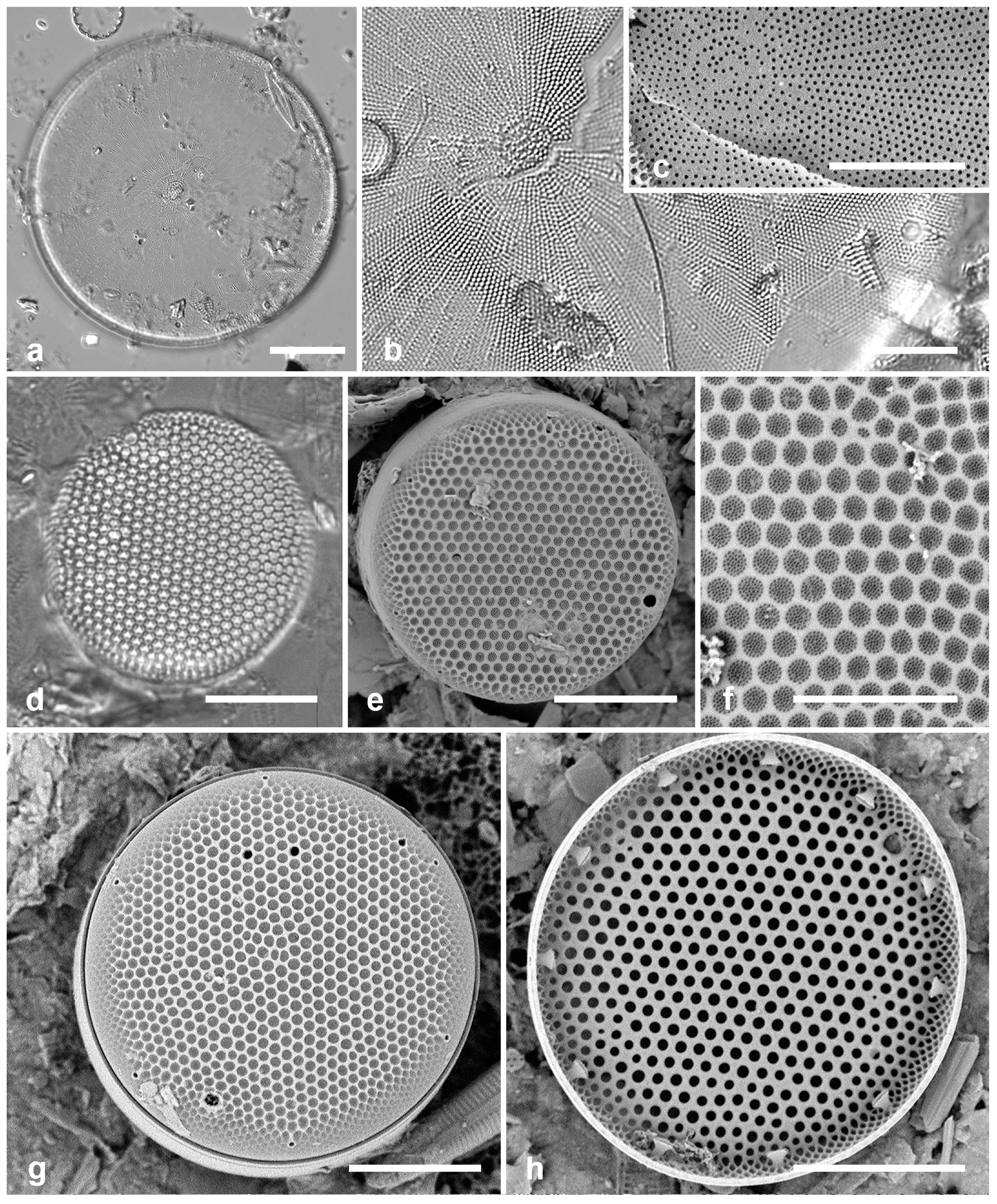
Diagnostics: Large valves with interfascicular rows indistinct and central area 7 µm with many areolae and a hyaline boundary.
3.7. Roperia cf. tesselata (Roper) Grunow ex Pelletan 1889 [51]—Figure 4d–h
Yap samples: Y18B, Y26C, Y34B, E, F, Y42-1
Dimensions: Diam. 20–30 µm, areolae 9–10 in 10 µm.
ASTEROLAMPRALES Round & R.M. Crawford
Asterolampraceae H.L. Smith
3.8. Asterolampra marylandica Ehrenberg 1844 [54]—Figure 5a
Yap samples: Y42-1
Dimensions: Diam. 49–131 µm.
Comments: Planktonic species.
Probosciaceae R.W. Jordan & Ligowski
3.9. Proboscia alata (Brightwell) Sunderström 1986 [55]—Figure 5b,c
Yap samples: Y45-5, Y45-2
Dimensions: Conical valves 8–10 µm diam. at margin.
3.10. Rhizosolenia imbricata Brightwell 1858 [56]—Figure 5d–h
Yap samples: Y25B
Dimensions: Diam. 14 µm
Diagnostics: Valve (calyptra) conical and asymmetrical, with a narrow spine (outer extension of rimoportula) that fits into a groove. Large areolae in both valve and girdle bands. Girdle bands in lateral rows.
(a) Asterolampra marylandica, valve in LM. (b,c) Proboscia alata valve and detail of tip, SEM. (d–h) Rhizosolenia imbricata. (d)Valve and several girdle bands in dorsiventral view, LM. (e) Girdle bands in lateral view, SEM. (f) Long fragment of frustule showing arrangement of girdle bands in lateral rows, SEM. (g,h) Details of valve (V) and attached girdle bands in lateral and slightly oblique view, SEM, showing the spine on one side (arrow in Figure 5g; out of view to left in Figure 5h) and the matching groove (arrow, Figure 5h), extending onto the girdle. Scale bars (f) = 25 µm, (a,b,d,f,g) = 10 µm, (c,e) = 5 µm.
(a) Asterolampra marylandica, valve in LM. (b,c) Proboscia alata valve and detail of tip, SEM. (d–h) Rhizosolenia imbricata. (d)Valve and several girdle bands in dorsiventral view, LM. (e) Girdle bands in lateral view, SEM. (f) Long fragment of frustule showing arrangement of girdle bands in lateral rows, SEM. (g,h) Details of valve (V) and attached girdle bands in lateral and slightly oblique view, SEM, showing the spine on one side (arrow in Figure 5g; out of view to left in Figure 5h) and the matching groove (arrow, Figure 5h), extending onto the girdle. Scale bars (f) = 25 µm, (a,b,d,f,g) = 10 µm, (c,e) = 5 µm.

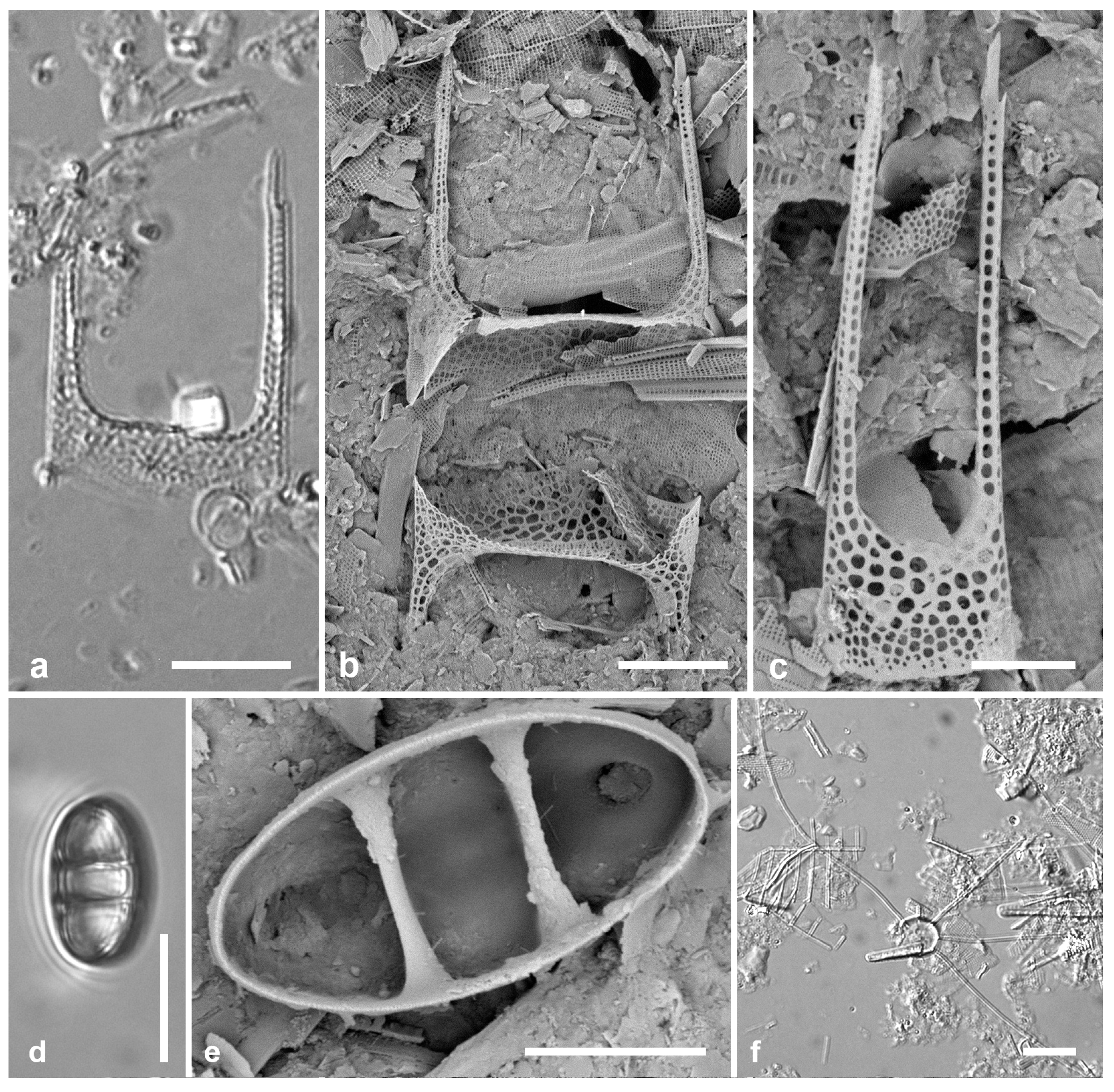
(a–c). Hemiaulus sinensis valves in girdle view in LM and SEM. (d,e) Anaulus minutus, SEM. (f) Bacteriastrum furcatum, LM. Scale bars: (a,b,d,f) = 10 µm, Figures (c,e) = 5 µm.
Figure 6.
(a–c). Hemiaulus sinensis valves in girdle view in LM and SEM. (d,e) Anaulus minutus, SEM. (f) Bacteriastrum furcatum, LM. Scale bars: (a,b,d,f) = 10 µm, Figures (c,e) = 5 µm.
HEMIAULALES Round & R.M. Crawford
3.11. Hemiaulus chinensis Greville 1865 [5]—Figure 6a–c
Yap samples: Y25B, Y26C
Dimensions: Length (apical axis) 10–25 µm.
Diagnostics: Valve elliptical, a coarse network of large areolae; two moderately long apical processes with a spine at the end (these used to connect the cells in chains).
ANAULALES Round & R.M. Crawford
Anaulaceae (Schütt) Lemmermann
3.12. Anaulus minutus Grunow in Van Heurck 1882 [44]—Figure 6d,e
Yap samples: Y26C, Y33A
Dimensions: Length 14 µm, width 8 µm
Diagnostics: Tiny elliptical valves divided by two pseudosepta, no evident areolae but a small elevation near each pole with opening.
CHAETOCEROTALES Round & R.M. Crawford
Chaetocerotaceae Ralfs in Prichard
3.13. Bacteriastrum furcatum Shadbolt 1853 [59]—Figure 6f
Yap samples: Y26B
Dimensions: Diam. of disc 8–11 µm
Comments: Planktonic. The setae typically fork once, though this is not apparent in the LM image and, in SEM images from Yap, setae were all broken off near the base. Specimens from other islands show this.
3.14. Chaetoceros peruvianus Brightwell 1856 [60]—Figure 7a–d
Yap samples: Y26B, Y26C
Dimensions: Diam. 18–24 µm
THALASSIOSIRALES Glezer & Makarova
Lauderiaceae (Schütt) Lemmermann emend. Lobban
3.15. Disymmetria excentrica (Lobban) Lobban 2023 [61]—Figure 7e
Synonym: Lauderia excentrica Lobban
Yap samples: Y41-8
Dimensions: 26 µm diameter, striae (measured by the costae below the reniform area) 15 in 10 µm.
Diagnostics: Discoidal, bipolar valves with numerous fultoportulae around the periphery and across the valve face; a reniform area of scattered pores set in one half of the valve face with striae and costae radiating from it. Only the periphery is pseudoloculate.
3.16. Disymmetria reticulata Lobban 2023 [61]
Dimensions: 26 µm diam., striae 10 in 10 µm.
Diagnostics: Discoidal, bipolar valves with pseudoloculate structure and fultoportulae in both periphery and reniform area; periphery wider at one pole.
(a–d) Chaetoceros peruvianus. (a) Upper valve in LM, showing base of setae. (b) Upper valve in SEM, in girdle view, showing large external rimoportula tube (arrow) between recurved setae. (c,d) Lower valve with straight setae and detail of pores and spines on seta. (e) Disymmetria excentrica, SEM. Scale bars: (a–c) = 10 µm, (e) = 5 µm, (d) = 2 µm.
Figure 7.
(a–d) Chaetoceros peruvianus. (a) Upper valve in LM, showing base of setae. (b) Upper valve in SEM, in girdle view, showing large external rimoportula tube (arrow) between recurved setae. (c,d) Lower valve with straight setae and detail of pores and spines on seta. (e) Disymmetria excentrica, SEM. Scale bars: (a–c) = 10 µm, (e) = 5 µm, (d) = 2 µm.
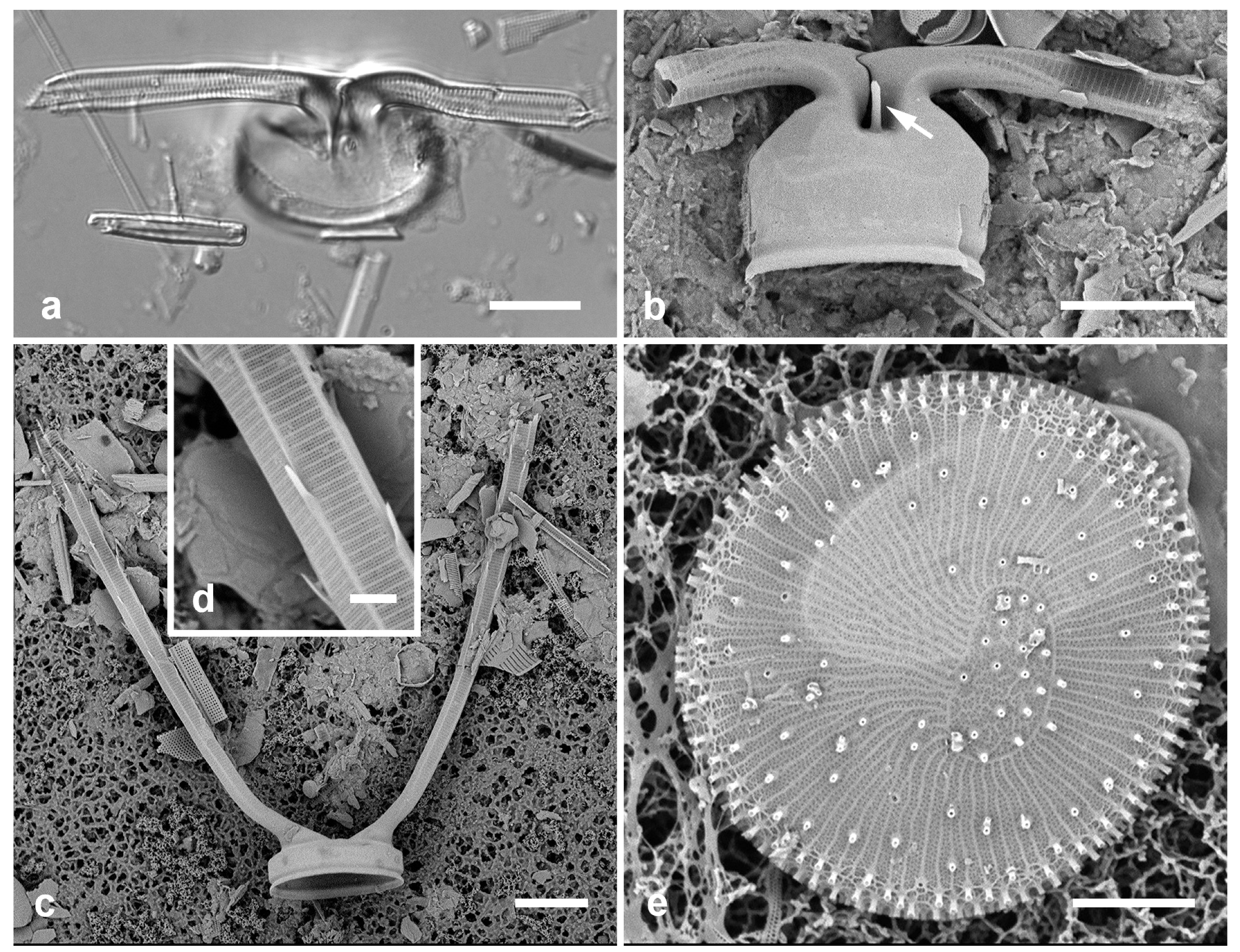
3.17. Skeletonema grevillei Sarno & Zingone 2005 [62]—Figure 8a–c
Yap samples: Y25B
Dimensions: Diam. 8–9 µm
CYMATOSIRALES Round & R.M. Crawford
Cymatosiraceae Hasle, Stosch & Syvertsen
(a–c) Skeletonema grevillei, SEM. (a) Portion of chain. (b) Two cells in chain connected by linked extensions of the fultoportulae, joined in the middle. (c) Oblique view of internal surface (center of valve missing), showing openings of the single rimoportula (arrow) and the ring of fultoportulae (arrowheads). (d–f) Unidentified Cymatosiraceae in LM and two views of a valve in SEM, (f) rotated and tilted 60°. Scale bars: (a) = 25 µm, (d) = 10 µm, (e,f) = 5 µm, (b,c) = 2 µm.
Figure 8.
(a–c) Skeletonema grevillei, SEM. (a) Portion of chain. (b) Two cells in chain connected by linked extensions of the fultoportulae, joined in the middle. (c) Oblique view of internal surface (center of valve missing), showing openings of the single rimoportula (arrow) and the ring of fultoportulae (arrowheads). (d–f) Unidentified Cymatosiraceae in LM and two views of a valve in SEM, (f) rotated and tilted 60°. Scale bars: (a) = 25 µm, (d) = 10 µm, (e,f) = 5 µm, (b,c) = 2 µm.
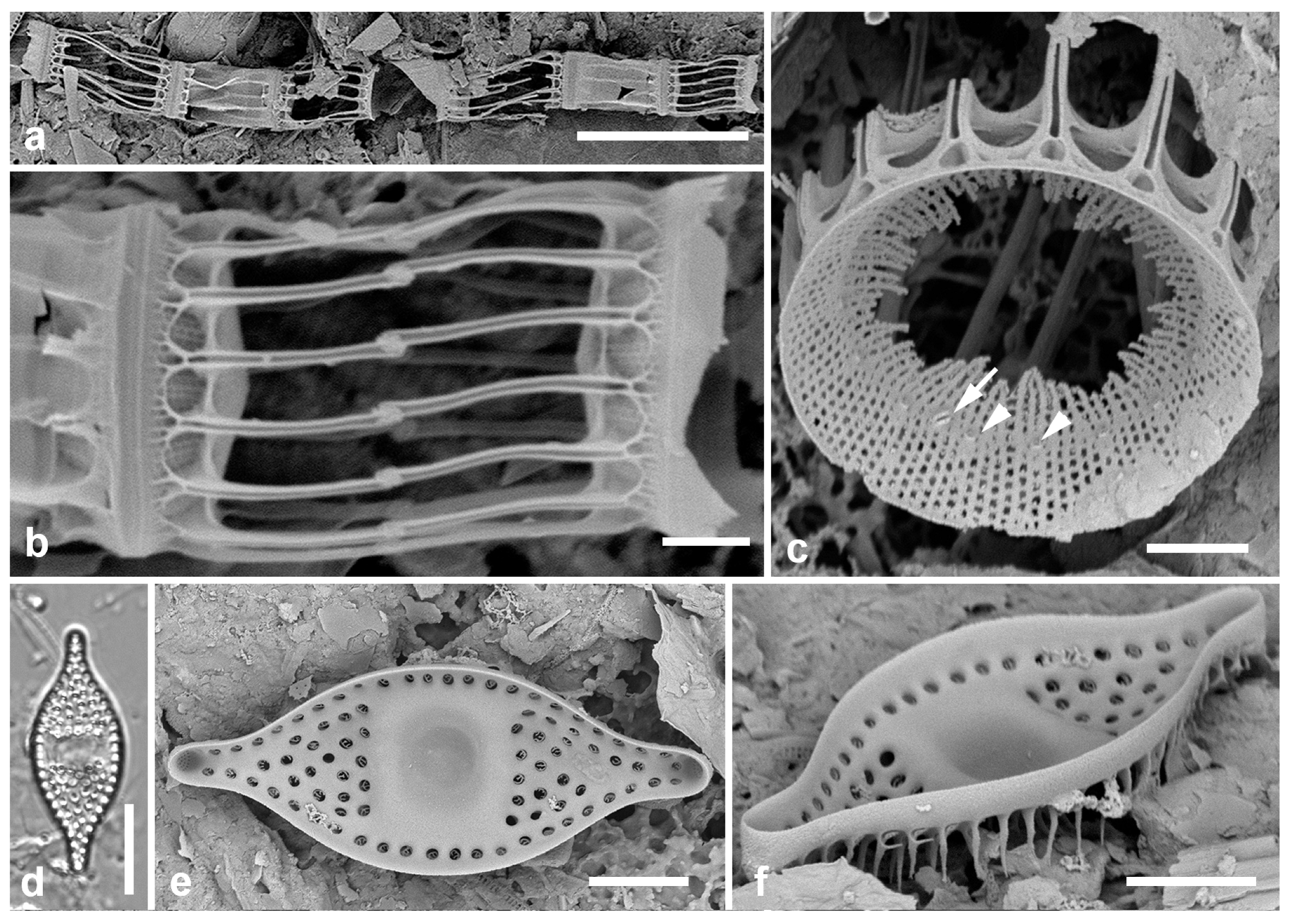
3.18. Unidentified Genus 1. Figure 8d–f
Yap samples: Y26C
Dimensions: Length 28 µm, width 10–11 µm
[EUPODISCALES E.J. Cox (invalid)]
Odontellacae P.A. Sims, D.M. Williams & Ashworth
3.19. Odontella obtusa Kützing 1844 [37]—Figure 9 a,b
Yap samples: Y16B
Dimensions: Length 33 µm (between ocelli).
(a,b). Odontella obtusa, SEM. (c,d) Pseudictyota dubia. (c) Valve in valve view, LM. (d) Valve in girdle view, SEM, showing ocelli (arrowhead), rimoportulae (arrow) and pseudoloculate structure. Scale bars: (a) = 25 µm, (b,c) = 10 µm, (d) = 5 µm.
Figure 9.
(a,b). Odontella obtusa, SEM. (c,d) Pseudictyota dubia. (c) Valve in valve view, LM. (d) Valve in girdle view, SEM, showing ocelli (arrowhead), rimoportulae (arrow) and pseudoloculate structure. Scale bars: (a) = 25 µm, (b,c) = 10 µm, (d) = 5 µm.
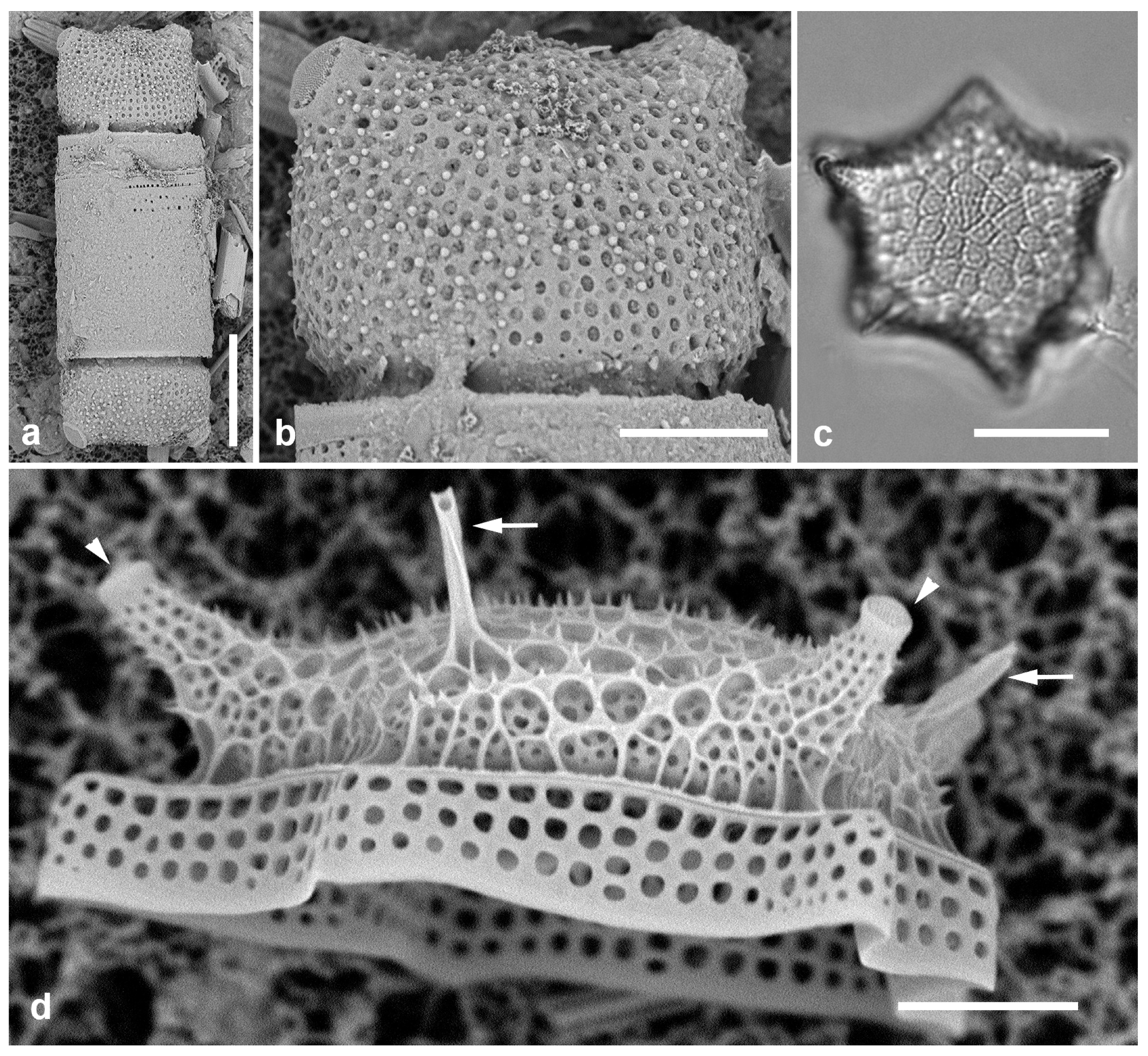
Diagnostics: Small cells with large areolae (not pseudoloculate—contrast Pseudictyota below), ocelli on low apical elevations (no higher than the central elevation).
3.20. Pseudictyota dubia (Brightwell) P.A.Sims & D.M.Williams 2018 in [66]—Figure 9c,d
Synonym: Triceratium dubium Brightwell
Yap samples: Y25H-1, Y25H-2, Y37-8, Y41-7
Dimensions: Diam. 24–27 µm.
ARDISSONEALES Round emend Lobban & Ashworth
Ardissoneaceae Round emend Lobban & Ashworth
3.21. Ardissonea densistriata Lobban 2022 in [23]
Additional Yap samples: Y41-1, Y42-3
Dimensions: Length 46–103 µm, width 7–10 µm, stria density 16–17 in 10 µm
Comments: Stria density nearly double that of A. formosa. Ardissoneaceae are either known or expected to be epiphytic, often attached to seaweeds by stout mucilage pads.
3.22. Ardissonea formosa (Hantzsch) Grunow 1880 in [68]
Additional Yap samples: Y41-7
Dimensions: Length 201–300 µm, width 22–24 µm, stria density 8–10 in 10 µm
3.23. Ardissoneopsis appressata Lobban & Ashworth 2022 in [23]
Additional Yap samples: Y36-1
Dimensions: Length 857 µm (560–900 µm), width 9–12 µm, striae 18–19 in 10 µm
3.24. Ardissoneopsis gracilis Lobban 2022 in [23]—Figure 10a,b
Additional Yap samples: Y26C
Dimensions: Length 260 µm, width 7 µm at poles, 8 µm at center, stria density 11 in 10 µm
(a–f) Ardissoneopsis. (a,b) A. gracilis valve from Y36-2 in LM with central portion and one pole; detail of central portion, typical of the species. (c–f) Valve fragments from Y26C, possibly a different species but not A. undosa. (c) Mid portion with inflation, LM. (d) Internal SEM of pole, showing increased stria density at apex. (e) External view of apex, SEM, showing spines; broken edge shows lack of internal costae. (f) Internal aspect of central portion, SEM, showing location of annulus (arrow). Scale bars: (a) = 25 µm, (b–f) = 10 µm.
Figure 10.
(a–f) Ardissoneopsis. (a,b) A. gracilis valve from Y36-2 in LM with central portion and one pole; detail of central portion, typical of the species. (c–f) Valve fragments from Y26C, possibly a different species but not A. undosa. (c) Mid portion with inflation, LM. (d) Internal SEM of pole, showing increased stria density at apex. (e) External view of apex, SEM, showing spines; broken edge shows lack of internal costae. (f) Internal aspect of central portion, SEM, showing location of annulus (arrow). Scale bars: (a) = 25 µm, (b–f) = 10 µm.

3.25. Climacosphenia elegantissima Lobban 2022 in [23]—Figure 11a
Yap samples: Y26B, Y26C, Y41-8
Dimensions: Length > 1000 µm, width across apex 25 µm, striae 27–28 in 10 µm
Comments: Differing from C. elongata J.W. Bailey (also present in Micronesia) in the very long, narrow stem and margins and sides of the annulus parallel toward the apex.
3.26. Climacosphenia scimiter A. Mann 1925 [9]—Figure 11b
Yap samples: Y41-7, Y41-8
Dimensions: Length 273 µm, width across apex 27 µm, striae 29 in 10 µm
3.27. Grunowago pacifica Lobban & Ashworth 2022 in [23]—Figure 11c
Yap samples: Y42-1
Dimensions: Length 272 µm, width 13 µm, striae 8 in 10 µm
Comments: This species of “big sticks” differs from other Ardissoneaceae in having a longitudinal costa along the apical axis. The species differs from Grunowago bacillaris (Grunow) Lobban & Ashworth in width and lanceolate valve outline, not reported from Micronesia.
(a) Climacosphenia elegantissima, apical portion of valve and valvocopula showing parallel sides and annular lines until the larger space between craticular bars, LM. (b) Climacosphenia scimiter, valvocopula, LM. (c) Grunowago pacifica, SEM of valve interior and valvocopula, showing central costa. (d,e) Synedrosphenia gomphonema, LM. (f) Synedrosphenia licmophoropsis, apical and middle portions in LM, arrows indicate annulus. Scale bars: (a,b,d) = 25 µm, (c) = 20 µm, (e,f) = 10 µm.
Figure 11.
(a) Climacosphenia elegantissima, apical portion of valve and valvocopula showing parallel sides and annular lines until the larger space between craticular bars, LM. (b) Climacosphenia scimiter, valvocopula, LM. (c) Grunowago pacifica, SEM of valve interior and valvocopula, showing central costa. (d,e) Synedrosphenia gomphonema, LM. (f) Synedrosphenia licmophoropsis, apical and middle portions in LM, arrows indicate annulus. Scale bars: (a,b,d) = 25 µm, (c) = 20 µm, (e,f) = 10 µm.
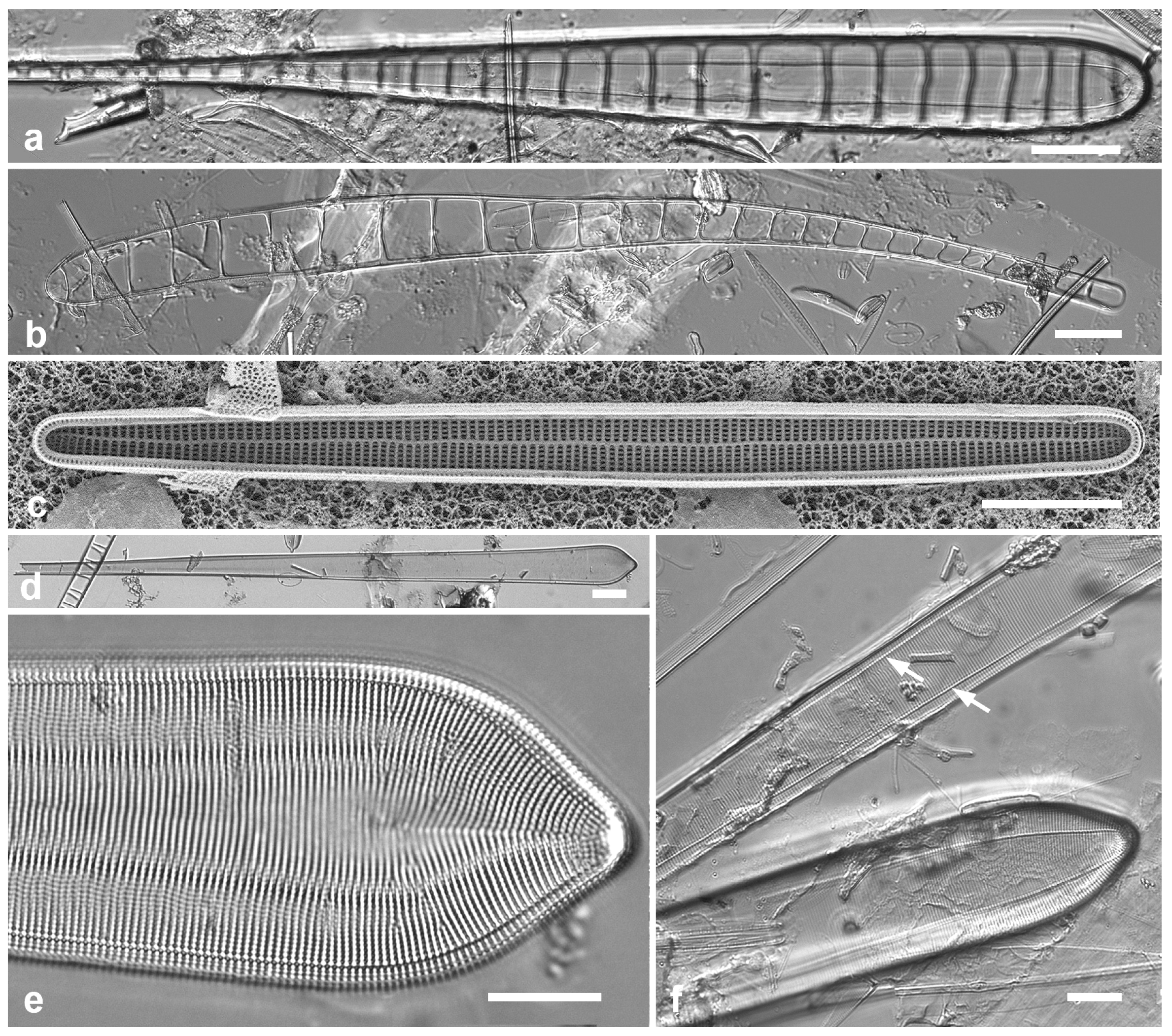
3.28. Synedrosphenia gomphonema (Janisch & Rabenhorst) Hustedt 1932 [69]—Figure 11d,e
Yap samples: Y42-1
Dimensions: Length 504 µm, width 29 µm.
3.29. Synedrosphenia licmophoropsis Lobban 2022 in [23]—Figure 11f
Yap samples: Y37-8
Dimensions: Length 600–735 µm, widening apically from 14 to 27 µm, striae 19 in 10 µm near apex
(a) Toxarium hennedyanum, central portion showing smooth valve outline and field of scattered areolae inside the annulus; LM. (b) Toxarium cf. hennedyanum central and apical portions of a valve with no areolae inside the annulus (annulus along the valve margin with a line of areolae on each side); LM. (c) Toxarium undulatum, center and apical portions of valves showing undulating outline and scattered areolae inside the annulus at center and pole; SEM. Scale bars = 10 µm.
Figure 12.
(a) Toxarium hennedyanum, central portion showing smooth valve outline and field of scattered areolae inside the annulus; LM. (b) Toxarium cf. hennedyanum central and apical portions of a valve with no areolae inside the annulus (annulus along the valve margin with a line of areolae on each side); LM. (c) Toxarium undulatum, center and apical portions of valves showing undulating outline and scattered areolae inside the annulus at center and pole; SEM. Scale bars = 10 µm.
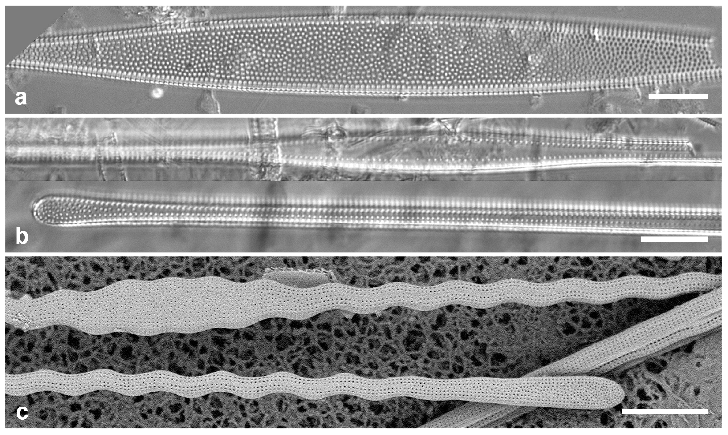
3.30. Toxarium hennedyanum (Gregory) Pelletan 1889 [51]—Figure 12a,b
Yap samples: Y26B, Y26C
Dimensions: Length 156–334 µm in Micronesia; width 7 µm across inflated center; striae 9–11 in 10 µm
3.31. Toxarium undulatum Bailey ex Bailey 1854 [70]—Figure 12c
Yap samples: Y26C, Y36-2, Y42-3
Dimensions: Length 450 µm, width 6 µm, striae 14 in 10 µm
3.32. Biddulphia biddulphiana (J.E.Smith) Boyer 1901 [71]—Figure 13a
Yap samples: Y25H-2
Dimensions: Length 79 µm
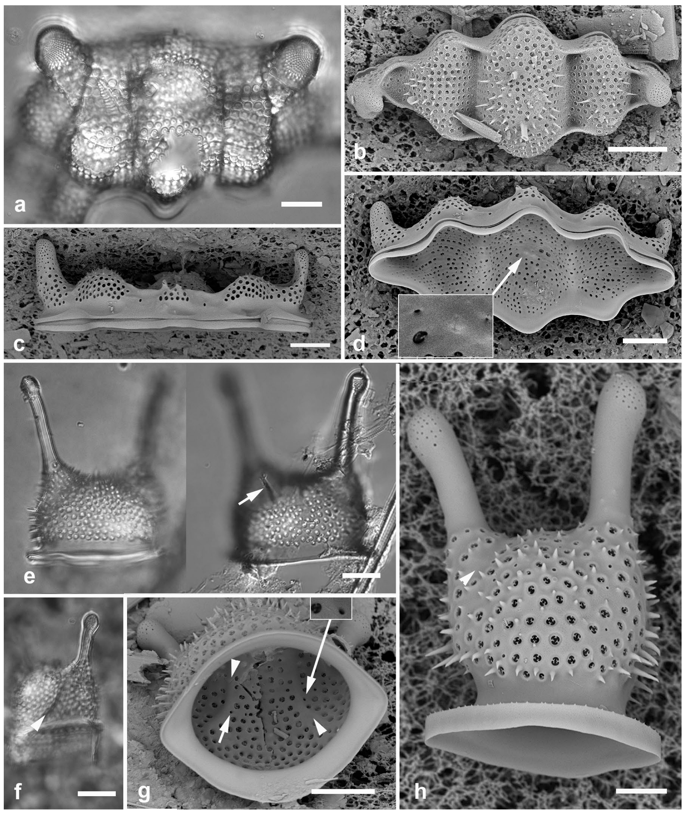
3.33. Biddulphiella cuniculopsis Lobban, sp. nov.—Figure 13e–h
Diagnostics: Distinguished from congeners by long apical elevations arising from a single elevation, in valve view elliptical–rhomboidal.
Type locality: GUAM: GabGab reef, Piti Municipality,Apra Harbor, 13.443 N, 144.643 E, associated with Halimeda and its epiphytes, ca. 10 m depth, sample GU44AR-3. Coll. C.S. Lobban and M. Schefter, 12 August 2012.
Additional records: Y 26C, Y45-2
Etymology: From L. cuniculus, a rabbit, with reference to the resemblance to a rabbit’s head and ears.
3.34. Biddulphiella tridens (Ehrenberg) P.A. Sims & M.P. Ashworth 2022 in [72]—Figure 13b–d
Synonym: Biddulphia tuomeyi (J.W. Bailey) Roper
Yap samples: Y26C, Y34E
Dimensions: Length 55–70 µm, width 23–28 µm.
(a) Biddulpha biddulphiana, oblique valve in LM. (b–d) Biddulphiella tridens, SEM. (b) Exterior view showing deep sulci, spines, and two rimoportula tubules (arrows). (c) Valve in profile, tilt = 80°. (d) Valve as in (c), tilt = 0°, inset showing internal rimoportula opening. (e–h) Biddulphiella cuniculopsis, n. sp. (e) LM holotype valve from Guam at two focal planes in near-apical girdle view, showing rimoportula (arrow). (f) LM specimen from Yap, showing sulcus (arrowhead). (g,h) SEM of valve from Yap. (g) Interior, tilt = 40°, showing sulci (arrowheads) and possible rimoportula opening (arrows; compare Figure 13e). (h) Valve in lateral girdle view, showing typical shape; arrowhead = sulcus. Scale bars: (a–g) = 10 µm, (h) = 5 µm.
(a) Biddulpha biddulphiana, oblique valve in LM. (b–d) Biddulphiella tridens, SEM. (b) Exterior view showing deep sulci, spines, and two rimoportula tubules (arrows). (c) Valve in profile, tilt = 80°. (d) Valve as in (c), tilt = 0°, inset showing internal rimoportula opening. (e–h) Biddulphiella cuniculopsis, n. sp. (e) LM holotype valve from Guam at two focal planes in near-apical girdle view, showing rimoportula (arrow). (f) LM specimen from Yap, showing sulcus (arrowhead). (g,h) SEM of valve from Yap. (g) Interior, tilt = 40°, showing sulci (arrowheads) and possible rimoportula opening (arrows; compare Figure 13e). (h) Valve in lateral girdle view, showing typical shape; arrowhead = sulcus. Scale bars: (a–g) = 10 µm, (h) = 5 µm.
(a–c) Biddulphiopsis membranacea, LM. (a,b). Complete valve (edges indicated by arrowheads) and detail of center showing scattered pattern contrasting with radiating striae. (c) Copula with apical septa. (d,e) Lampriscus shadboltianus. Valves in valve and girdle view, respectively, SEM, showing the smooth outline of the nominate variety and the characteristic spines on the edges of the ocelli in this species (arrow). Scale bars: (a,c) = 25 µm, (b,d,e) = 10 µm.
Figure 14.
(a–c) Biddulphiopsis membranacea, LM. (a,b). Complete valve (edges indicated by arrowheads) and detail of center showing scattered pattern contrasting with radiating striae. (c) Copula with apical septa. (d,e) Lampriscus shadboltianus. Valves in valve and girdle view, respectively, SEM, showing the smooth outline of the nominate variety and the characteristic spines on the edges of the ocelli in this species (arrow). Scale bars: (a,c) = 25 µm, (b,d,e) = 10 µm.
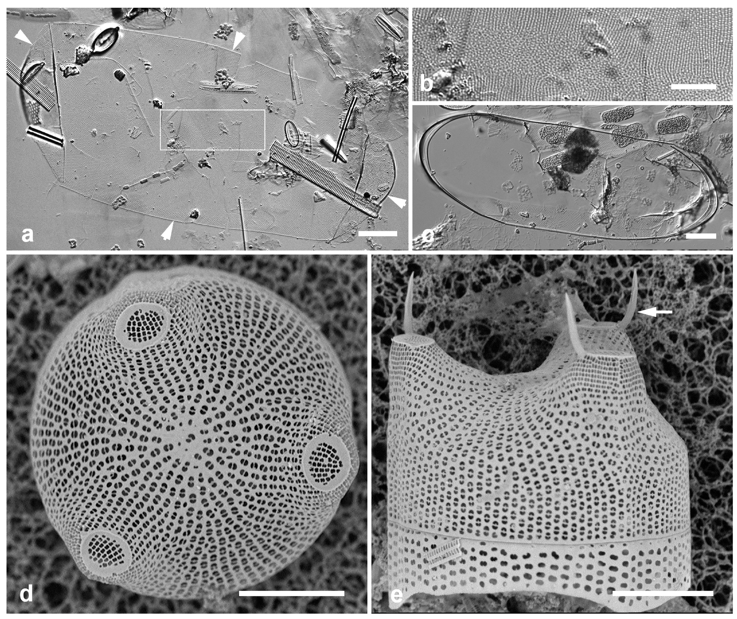
3.35. Biddulphiopsis membranacea (Cleve) von Stosch & Simonsen 1984 [74]—Figure 14a–c
Yap samples: Y42-1
Dimensions: Length 257–288 µm, width including mantle 121 µm, copula 100 µm.
3.36. Lampriscus shadboltianus (Greville) Peragallo & Peragallo 1902 [75]—Figure 14d,e
Yap samples: Y42-1
Dimensions: Diam. 29–31 µm
3.37. Striatella unipunctata (Lyngbye) Agardh 1832 [76]—Figure 15a
Yap samples: Y36-2, Y41-7, Y26C
PLAGIOGRAMMALES E.J. Cox (“nom. prov.”)
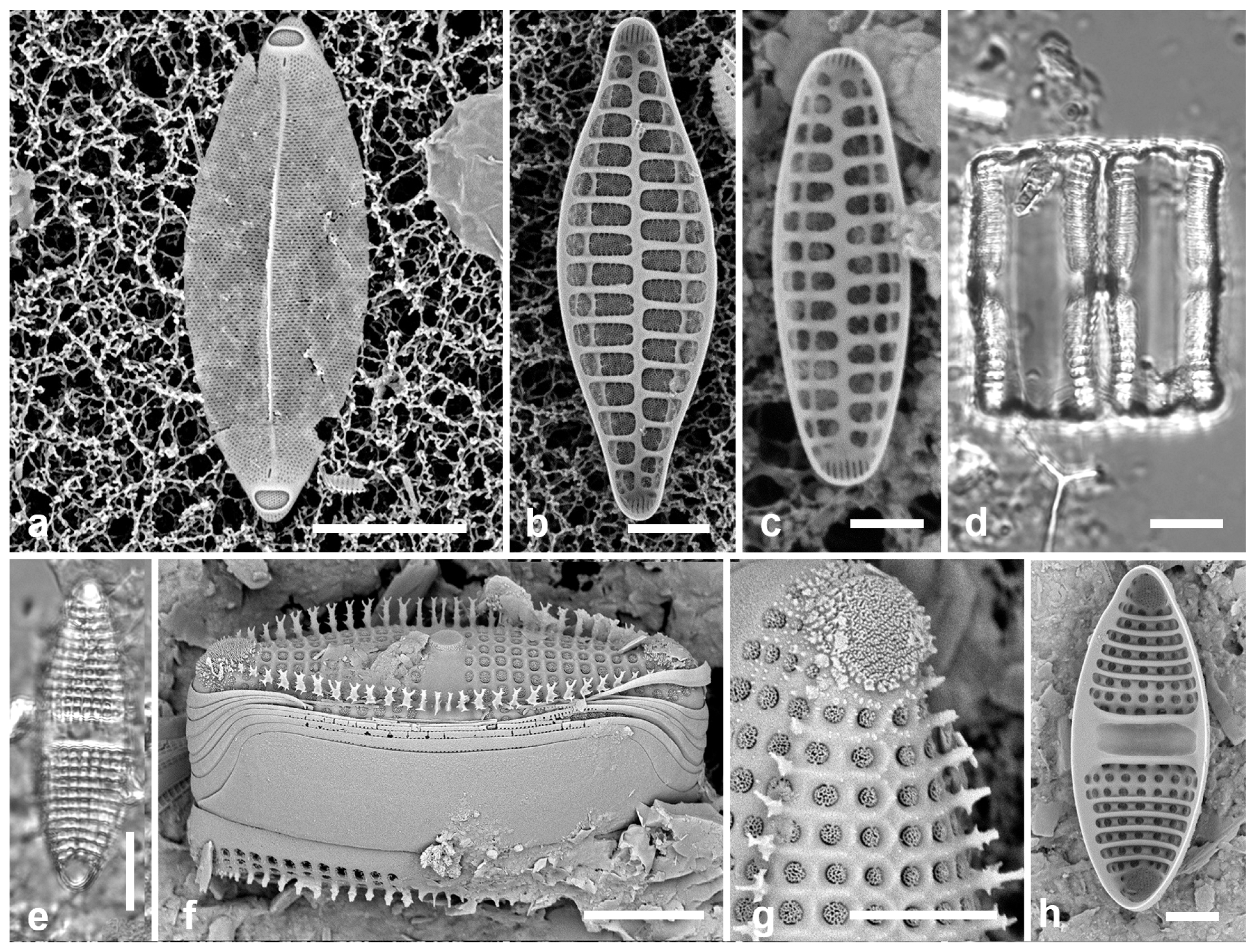
3.38. Neofragilaria anomala (Giffen) Witkowski & Dąbek 2015 in [77]—Figure 15b,c
Yap samples: Y16B, Y37-8, Y36-1, Y41-7, -8
Dimensions: Length 12–32 µm, width 4–10 µm, striae 5.5–12.5
3.39. Plagiogramma porcipellis Ashworth & Chunlian Li 2020 in [78]—Figure 15d–h
Yap samples: Y26C
Dimensions: Length 33–45 µm, width 13 µm; 8 striae in 10 µm.
3.40. Plagiogramma subatomus Lobban, S. Konno, Y. Arai & R.W. Jordan 2021 in [21]
Additional Yap samples: Y36-4
Dimensions: Length 9 µm, width 5 µm, areolae ca. 17 in 10 µm
(a) Striatella unipunctata, SEM. (b,c) Neofragilaria anomala, internal views of two valves. (d–h). Plagiogramma porcipellis. (d) Two frustules in girdle view, LM. (e) Valve in LM. (f) Frustule in oblique view, SEM, showing spines, apical pores fields, central elevation, and broad valvocopula. (g) Detail of apical pore field and areolae, SEM. (h) Internal view of valve showing pseudoseptum and transverse costae. Scale bars: (a,d,e,f,h) = 10 µm, (b,g) = 5 µm. (c) = 2 µm.
Figure 15.
(a) Striatella unipunctata, SEM. (b,c) Neofragilaria anomala, internal views of two valves. (d–h). Plagiogramma porcipellis. (d) Two frustules in girdle view, LM. (e) Valve in LM. (f) Frustule in oblique view, SEM, showing spines, apical pores fields, central elevation, and broad valvocopula. (g) Detail of apical pore field and areolae, SEM. (h) Internal view of valve showing pseudoseptum and transverse costae. Scale bars: (a,d,e,f,h) = 10 µm, (b,g) = 5 µm. (c) = 2 µm.
Psammodiscaceae Round & D.G. Mann
3.41. Psammodiscus nitidus (W.Gregory) Round & D.G. Mann 1980 [80]—Figure 16a,b
Yap samples: Y26C, Y34A
Dimensions: Diam. 17–21 µm
Diagnostics: Circular with large areolae. In SEM areolae covered with rotae supported by 2–3 spokes, presence of central pore, and usually one or more marginal or central rimoportulae.
(a,b) Psammodiscus nitidus in LM and SEM external view. (c) Rhaphoneis castracanei, LM. (d) Perissonoë crucifera, SEM external view. (e) Bleakeleya notata, LM. (f) Perideraion montgomeryi, SEM, external. (g,h) Falcula paracelsiana, SEM external, detail of apex with slits. Scale bars: (h) = 25 µm, (a,c–e) = 10 µm, (b,f,g) = 5 µm.
Figure 16.
(a,b) Psammodiscus nitidus in LM and SEM external view. (c) Rhaphoneis castracanei, LM. (d) Perissonoë crucifera, SEM external view. (e) Bleakeleya notata, LM. (f) Perideraion montgomeryi, SEM, external. (g,h) Falcula paracelsiana, SEM external, detail of apex with slits. Scale bars: (h) = 25 µm, (a,c–e) = 10 µm, (b,f,g) = 5 µm.
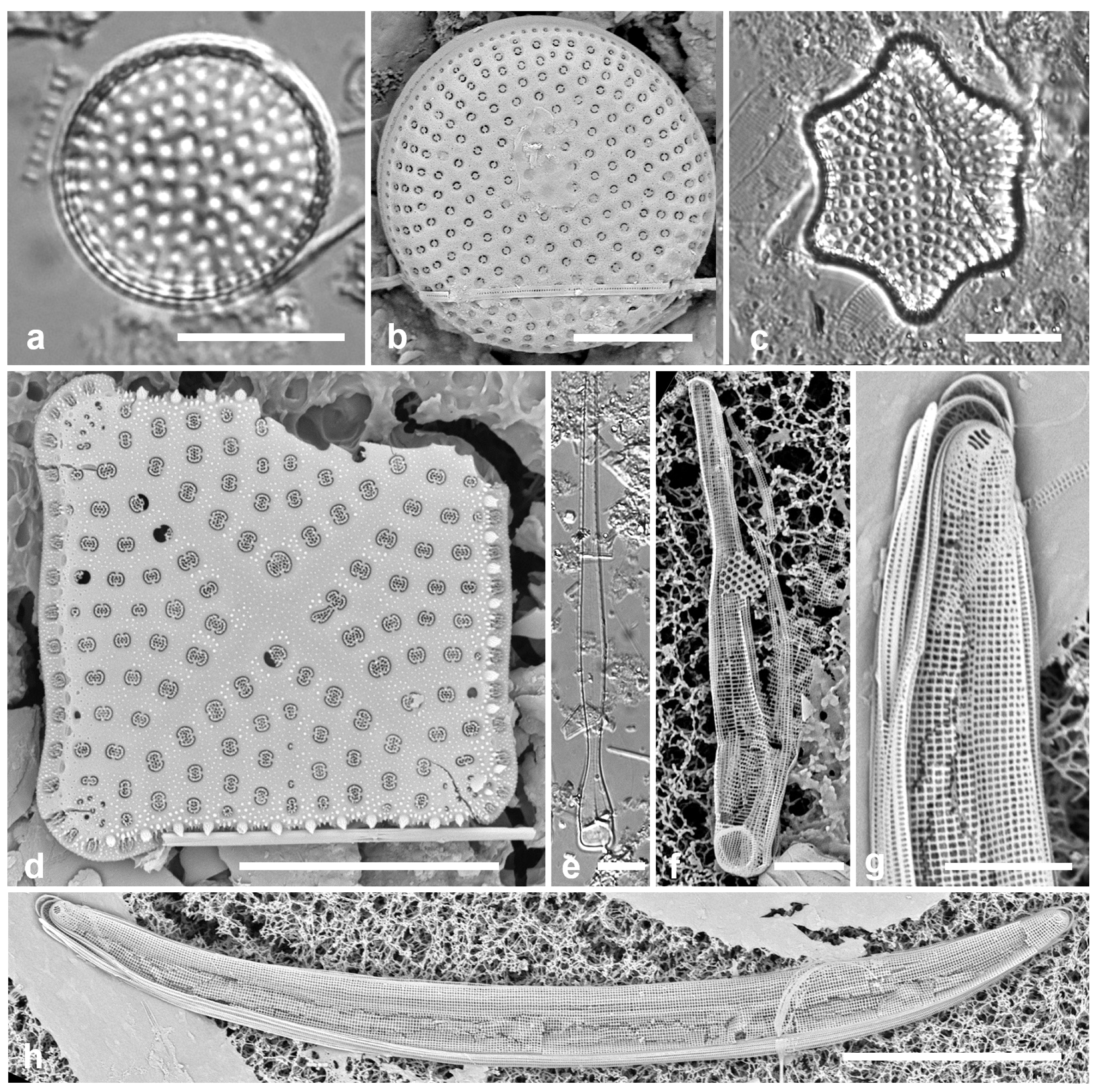
3.42. Perissonoë crucifera (Kitton in Prichard) Desikachary, Gowthaman, Hema, A.K.S.K. Prasad & Prema 1987 [82]—Figure 16d
Synonym: Perissonoë cruciata (Janisch & Rabenhorst) G.W. Andrews & Stoelzel
Yap samples: Y26C
Dimensions: Width 20 µm
3.43. Rhaphoneis castracanei Grunow in Van Heurck 1880 [44]—Figure 16c
Yap samples: Y26C
KOERNERELLALES Lobban & Ashworth
Koernerellaceae Lobban & Ashworth
3.44. Bleakeleya notata (Grunow in Van Heurck) F.E. Round 1990 in [52]—Figure 16e
Yap samples: Y36-1, Y41-7
Dimensions: Length 91 µm, width at base 9 µm
Comments: Loosely epiphytic, the necklace-like chains of this and Perideraion spp. loosely festooning seaweeds in sheltered waters.
3.45. Perideraion montgomeryi Lobban, Jordan & Ashworth 2011 in [85]—Figure 16f
Yap samples: Y41-8
3.46. Divergita biformis Lobban 2021 [21]
Yap samples: Y45-5
Dimensions: Length 76 µm, width 4 µm, striae 26–28 in 10 µm.
3.47. Falcula paracelsiana Voigt 1961 [86]—Figure 16g,h
Yap samples: Y45-2
Dimensions: Length 118 µm, striae 32 in 10 µm.
3.48. Hendeyella lineata Ashworth & Lobban 2016 in [89]—Figure 17a,b
Yap samples: Y37-8
Dimensions: Length 37 µm, width 5 µm, striae 10 in 10 µm
Diagnostics: Chains of linear valves tightly linked by stout spines.
Hendeyella. (a,b) Hendeyella lineata in SEM. (a) Yap voucher (Y37-8). (b) Guam specimen (GU44BF-1A) showing broad spines (arrowhead), broad valvocopula (VC), ligulate copula (arrow) and apical pore fields. (c–f) Hendeyella rhombica. (c) Chain in girdle view with valve view, LM. (d) Chain in girdle view showing broad valvocopula (VC) and narrowly branched spines, SEM. (e,f) Valve interiors, SEM, the latter showing weak heteropolarity, SEM. Scale bars: (a,c) = 10 µm, (b,d–f) = 5 µm.
Figure 17.
Hendeyella. (a,b) Hendeyella lineata in SEM. (a) Yap voucher (Y37-8). (b) Guam specimen (GU44BF-1A) showing broad spines (arrowhead), broad valvocopula (VC), ligulate copula (arrow) and apical pore fields. (c–f) Hendeyella rhombica. (c) Chain in girdle view with valve view, LM. (d) Chain in girdle view showing broad valvocopula (VC) and narrowly branched spines, SEM. (e,f) Valve interiors, SEM, the latter showing weak heteropolarity, SEM. Scale bars: (a,c) = 10 µm, (b,d–f) = 5 µm.
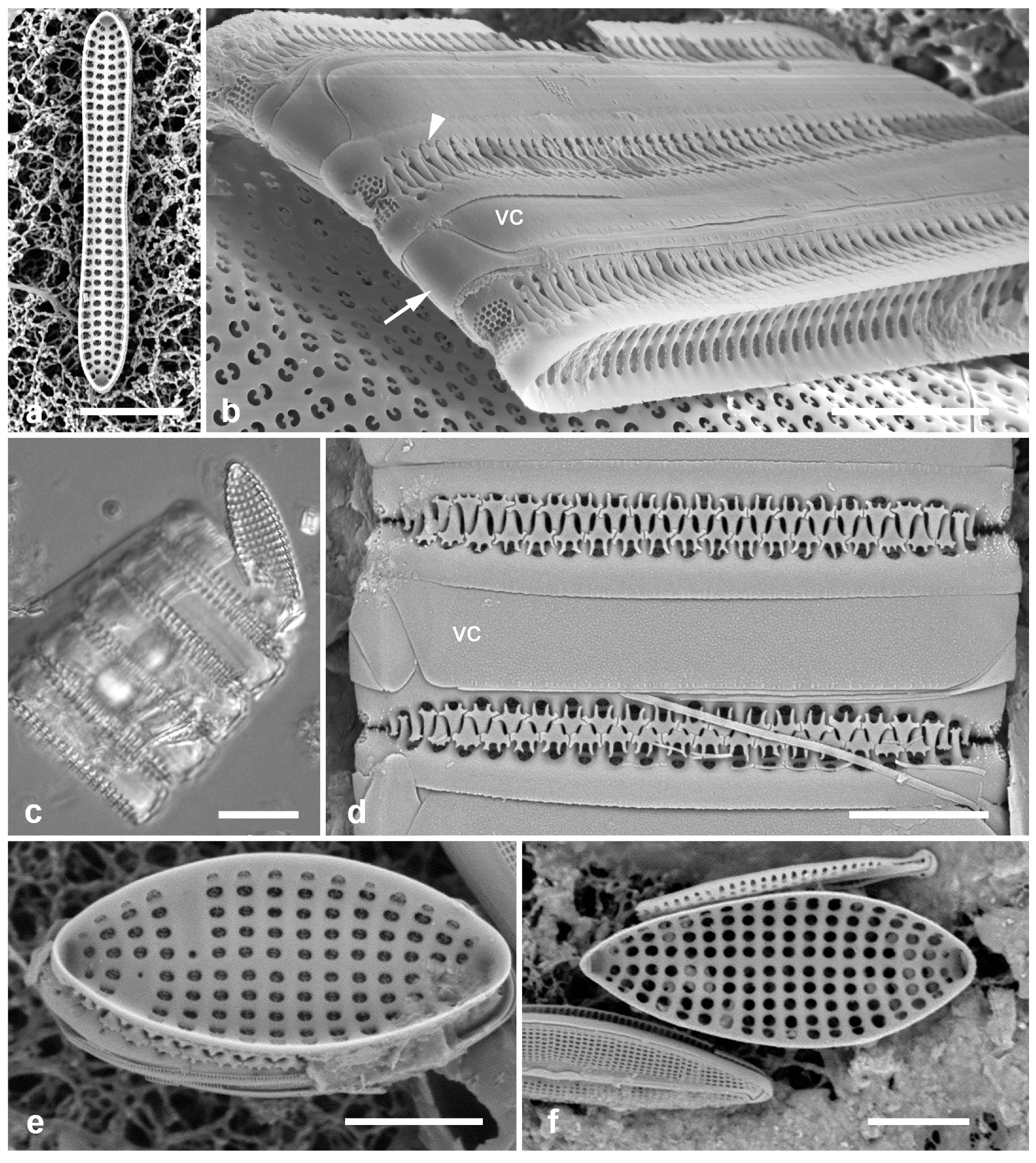
3.49. Hendeyella rhombica Ashworth 2016 in [89]—Figure 17c–f
Yap samples: Y18E, Y26C, Y37-8
Dimensions: Length 17–26 µm, width 7–8 µm, striae 10 in 10 µm
Diagnostics: Broadly lanceolate, slightly heteropolar valves tightly linked in chains with slender branched spines.
Comments: Valves are similar to H. dimeregrammopsis Ashworth, which is smaller (7.5–12 µm long, 4.4–5.8 µm wide) and less elliptical; on the basis of size, we have assigned the specimens in valve view and the LM image to H. rhombica. The girdle view in SEM shows the distinctively slender spines of this species. New record for Micronesia.
3.50. Hyalosynedra laevigata (Grunow) D.M. Williams & F.E. Round 1986 [90]—Figure 18a–f
Yap samples: Y41-7, Y45-5
Dimensions: Length 53–195 µm, width 4–5 µm, striae 52 in 10 µm
Diagnostics: Narrowly lanceolate, hyaline cells, distinguished from Stricosus cardinalii Lobban & E.C. Theriot in SEM by only three rows of pores in ocellulimbus and asymmetrical halves of the narrow rimoportula.
3.51. Stricosus cardinalii Lobban & E.C. Theriot 2018 in [91]—Figure 18f,g
Yap samples: Y16B
Dimensions: Length 107–116 µm, width 6 µm; striae (from literature) 38–39 in 10 µm
Diagnostics: Narrowly lanceolate, hyaline cells, distinguished from Hyalosynedra laevigata in SEM by 6–7 rows of pores in ocellulimbus and symmetrical halves of the wide rimoportula.
Comments: See Hyalosynedra laevigata, above.
3.52. Stricosus harrisonii Lobban & E.C.Theriot 2018 in [91]—Figure 18h,i
Yap samples: Y18E, Y33A
Dimensions: Length 269 µm, width 7 µm; striae (from literature) 38–41 in 10 µm
Diagnostics: Very long, linear, hyaline valve; sternum and rimoportula visible in LM.
Hyalosynedra vs. Stricosus. (a–f) Hyalosynedra laevigata. (a) Valve in LM (Y41-7). (b) Fractured frustule in SEM (Y41-7), left-hand apex out of frame, shown in Figure 18d. (c,d) Internal view of valve fragment (Y45-5) and detail of apex showing asymmetrical rimoportula (arrow). (e) Detail of apex external showing shallow ocellulimbus (arrow) and apical spines. (f,g) Stricosus cardinalii apex internal detail showing symmetrical rimoportula (arrow), and whole of same valve showing similarity of size and shape to H. laevigata. (h,i) Stricosus harrisonii, LM. Scale bars: (h) = 25 µm, (a–c,g,i) = 10 µm, (d–f) = 2 µm.
Hyalosynedra vs. Stricosus. (a–f) Hyalosynedra laevigata. (a) Valve in LM (Y41-7). (b) Fractured frustule in SEM (Y41-7), left-hand apex out of frame, shown in Figure 18d. (c,d) Internal view of valve fragment (Y45-5) and detail of apex showing asymmetrical rimoportula (arrow). (e) Detail of apex external showing shallow ocellulimbus (arrow) and apical spines. (f,g) Stricosus cardinalii apex internal detail showing symmetrical rimoportula (arrow), and whole of same valve showing similarity of size and shape to H. laevigata. (h,i) Stricosus harrisonii, LM. Scale bars: (h) = 25 µm, (a–c,g,i) = 10 µm, (d–f) = 2 µm.
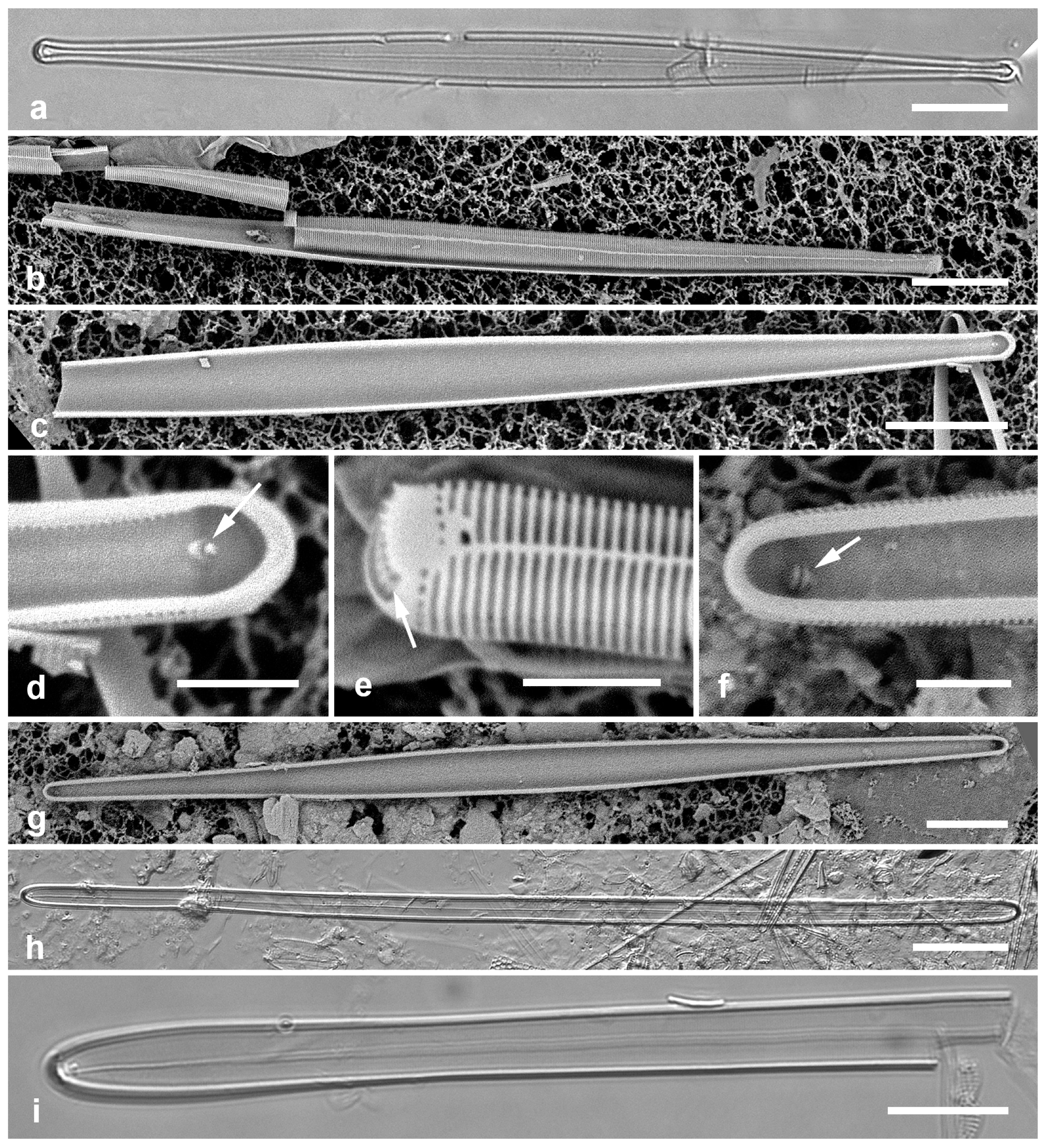
3.53. Neosynedra provincialis (Grunow) D.M. Williams & F.E. Round 1986 [90]—Figure 19a,b
Yap samples: Y45-5
Dimensions: Length 59 µm, width 3 µm.
(a,b) Neosynedra provincialis valve and detail of apex, SEM. (c,d). Neosynedra tortosa apex and valve, SEM. (e–h). Opephora pacifica. (e) Frustule in LM. (f,g) Valve interiors in SEM, showing size range and heteropolarity. (h) Apex external view showing pore field, SEM. (i,j) Synedra lata, SEM. Scale bars: (a,d–f,i,j) = 10 µm, (b,c) = 5 µm, (h) = 2 µm.
Figure 19.
(a,b) Neosynedra provincialis valve and detail of apex, SEM. (c,d). Neosynedra tortosa apex and valve, SEM. (e–h). Opephora pacifica. (e) Frustule in LM. (f,g) Valve interiors in SEM, showing size range and heteropolarity. (h) Apex external view showing pore field, SEM. (i,j) Synedra lata, SEM. Scale bars: (a,d–f,i,j) = 10 µm, (b,c) = 5 µm, (h) = 2 µm.
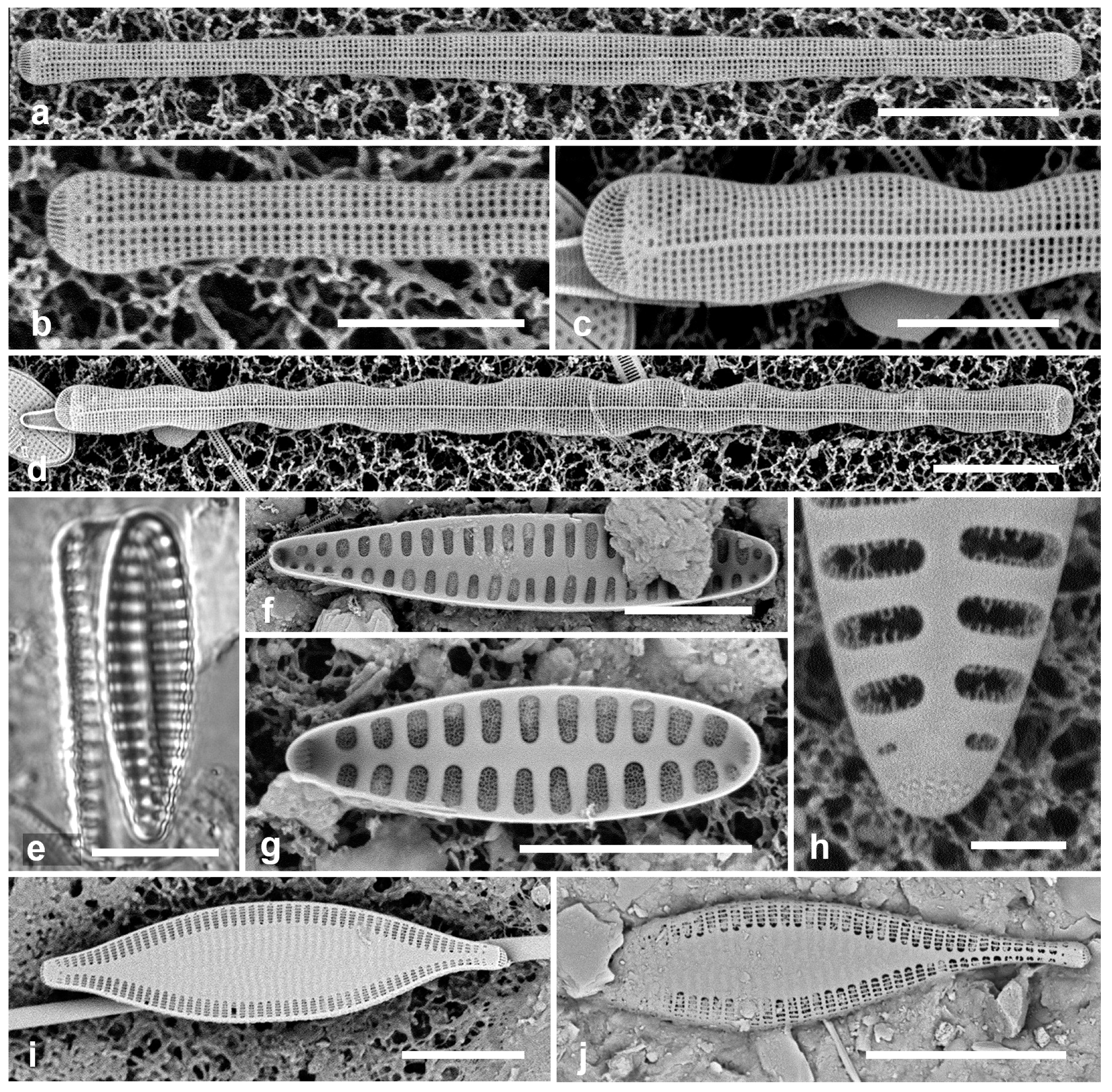
3.54. Neosynedra tortuosa (Grunow) D.M. Williams & F.E. Round 1986 [90]—Figure 19c,d
Additional Yap samples: Y25H-1, Y41-7
Dimensions: Length 85 µm, width 5 µm.
3.55. Synedra lata (Giffen) Witkowski 2000 in [46]—Figure 19i,j
Yap samples: Y37-5, Y37-8, Y26C
Dimensions: Length 30–40 µm, width 6–10 µm, striae 14–17 in 10 µm
Diagnostics: Broadly lanceolate, rostrate valves with short, coarse striae only along the margin, rest of valve surface hyaline. Apical pore fields apparently not sunken.
3.56. Tabularia parva (Kützing) D.M. Williams & F.E. Round 1986 [90]—Figure 20a–d
Yap samples: Y16B, Y42-1
Dimensions: Length 13–52 µm, width 4–5 µm, striae 20–22 in 10 µm
Diagnostics: Lanceolate cells with small apical ocellulimbi; broad, lanceolate sternum; biseriate striae on valve face and mantle with no break, areolae opposite; single rimoportula.
3.57. Opephora pacifica (Grunow) Petit 1899 [95]—Figure 19e–h
Yap samples: Y16B
Dimensions: Length 20–44 µm, width 7–8 µm, striae 6–7 in 10 µm
RHABDONEMATALES Round & R.M. Crawford
Grammatophoraceae Lobban & Ashworth
3.58. Grammatophora angulosa Ehrenberg 1840 [97]—Figure 20e
Yap samples: Y26C
Dimensions: Length 15 µm
Comments: Scarce and so far always very small in Micronesia.
3.59. Grammatophora oceanica Ehrenberg 1840 [98]—Figure 20f,g
Yap samples: Y36-4
Dimensions: Length 33 µm, width 7 µm, striae 22 in 10 µm
Diagnostics: Valves linear-elliptical, not inflated in the middle; stria density 22–23 in 10 µm. Based on stria densities and the lack of inflation, these are G. oceanica rather than G. marina (Lyngbye) Kützing, but the two species, each with several named varieties, are very difficult to distinguish.
Comments: Forming epiphytic zig-zag chains, along with other species of Grammatophora.
(a–d) Tabularia parva, SEM. (a,b) Large specimen and detail of striae. (c,d) Small specimens, (c) oblique, showing lack of break in striae at valve–mantle junction. (e) Grammatophora angulosa, girdle view showing characteristically hooked septa, LM. (f,g) Grammatophora oceanica, LM, valve view and girdle view. (h) Hyalosira tropicalis, SEM. (i) Microtabella interrupta, SEM of frustule showing valve interior and copulae with septa. Scale bars: (a,e–g,i) = 10 µm, (c,h) = 5 µm, (b,d) = 2 µm.
Figure 20.
(a–d) Tabularia parva, SEM. (a,b) Large specimen and detail of striae. (c,d) Small specimens, (c) oblique, showing lack of break in striae at valve–mantle junction. (e) Grammatophora angulosa, girdle view showing characteristically hooked septa, LM. (f,g) Grammatophora oceanica, LM, valve view and girdle view. (h) Hyalosira tropicalis, SEM. (i) Microtabella interrupta, SEM of frustule showing valve interior and copulae with septa. Scale bars: (a,e–g,i) = 10 µm, (c,h) = 5 µm, (b,d) = 2 µm.
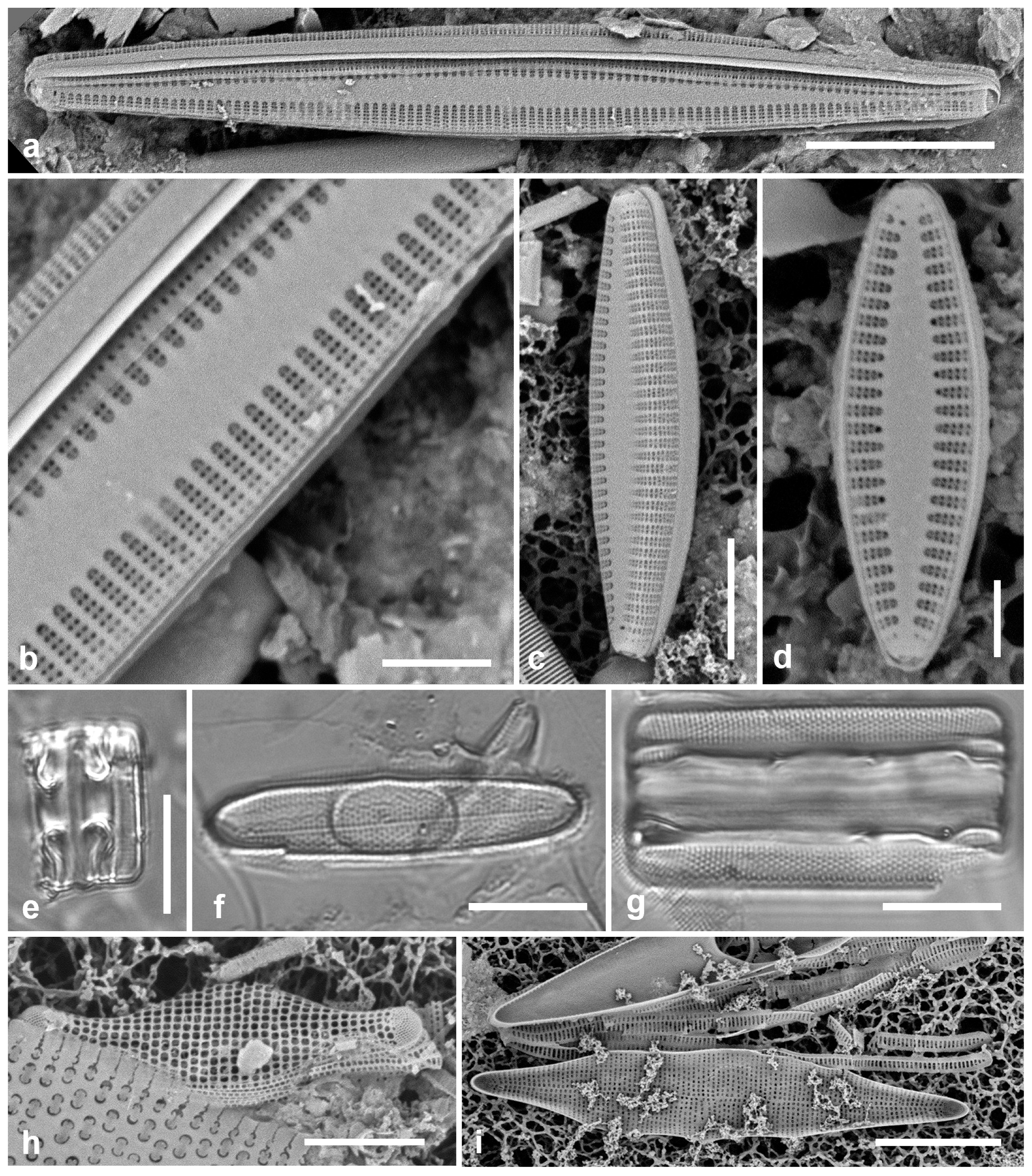
3.60. Hyalosira pacifica Lobban 2022 in [98]
Yap samples: Y26A
Dimensions: Length 9–23 µm, width 3–5 µm, striae 22–24 in 10 µm
Comment: Hyalosira pacifica type locality is Melbourne, Australia, attached to seaweeds by mucilage pads.
3.61. Hyalosira tropicalis J.N. Navarro 1992 in [101]—Figure 20h
Yap samples: Y41-8
Dimensions: Length 17 µm, width 5 µm, striae 24 in 10 µm
Diagnostics: Small, epiphytic, chain-forming cells, distinguished from congeners by the inflated valve outline and the uniseriate striae with large pores.
3.62. Microtabella interrupta (Ehrenberg) F.E. Round 1990 in [52]—Figure 20i and Figure 21a
Yap samples: Y25H-1, Y25H-2, Y41-7
Dimensions: Length 19–38 µm, width 7 µm, striae 26 in 10 µm
3.63. Microtabella rhombica Lobban 2015 [105]—Figure 21b,c
Yap samples: Y18E, Y26C
Dimensions: Length 37–52 µm, width 11–12 µm, striae 27–29 in 10 µm
Diagnostics: Rhombic valve much broader than M. interrupta but hard to distinguish in girdle view.
CYCLOPHORALES Round & R.M. Crawford
Cyclophoraceae Round & R.M. Crawford
3.64. Cyclophora minor Ashworth & Lobban 2012 in [106]—Figure 21d
Yap samples: Y41-8
Dimensions: Length 8 µm, striae 44 in 10 µm
Diagnostics: Very small lanceolate cells with circular pseudoseptum on one or both valves.
(a) Microtabella interrupta valve and girdle bands in LM. (b,c) Microtabella rhombica. (b) SEM, arrow points to apical spine. (c) Valve and girdle band in LM. (d) Cyclophora minor frustule in LM, one valve with pseudoseptum. (e) Cyclophora tenuis valve exterior in SEM, arrows indicate two parts of the apical slit field. (f) Licmophora flabellata valve in LM, showing multiple rimoportulae along sternum. Scale bars: (a–c,e,f) = 10 µm, (d) = 2 µm.
Figure 21.
(a) Microtabella interrupta valve and girdle bands in LM. (b,c) Microtabella rhombica. (b) SEM, arrow points to apical spine. (c) Valve and girdle band in LM. (d) Cyclophora minor frustule in LM, one valve with pseudoseptum. (e) Cyclophora tenuis valve exterior in SEM, arrows indicate two parts of the apical slit field. (f) Licmophora flabellata valve in LM, showing multiple rimoportulae along sternum. Scale bars: (a–c,e,f) = 10 µm, (d) = 2 µm.
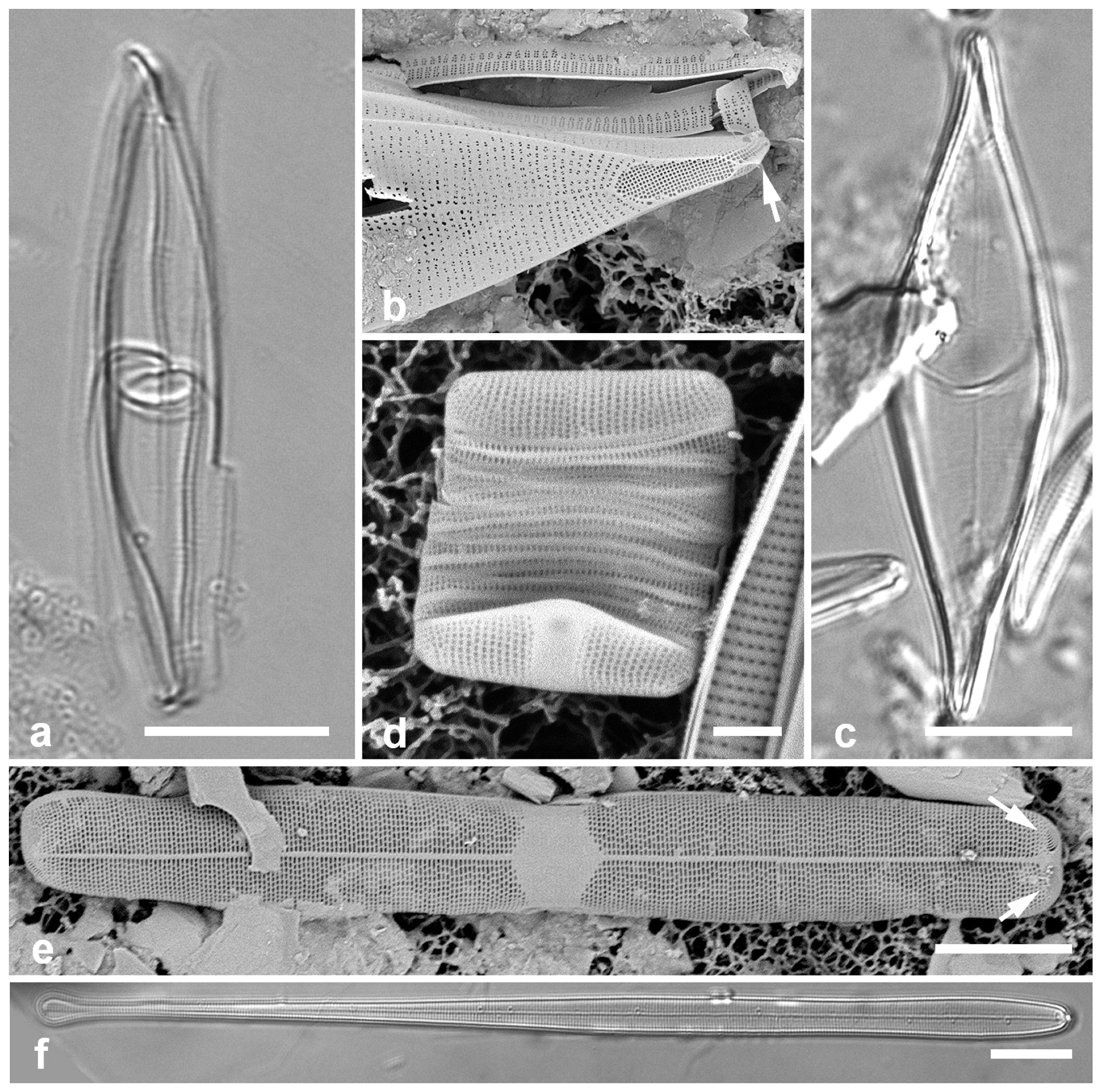
3.65. Cyclophora tenuis Castracane 1878 [107]—Figure 21e
Samples: Y34B
Dimensions: Length 76 µm, width 9 µm, striae 31 in 10 µm
Diagnostics: Broad valves, circular pseudoseptum on only one valve, apical slits in a U-shape field, often appearing as two patches in valve view.
3.66. Licmophora flabellata (Greville) C. Agardh 1831 [108]—Figure 21f
Yap samples: Y36-4, Y37-7
Dimensions: Length 60–150 µm, width 7–9 µm, striae 31–33
Comment: Well-known for attaching by strong mucilage stalks to seaweeds and other submerged surfaces, biofouling.
3.67. Licmophora cf. hastata Mereschkowsky 1901 [110]—Figure 22a–d
Yap samples: Y36-4, Y41-7
Dimensions: Length 38–50 µm, max. width 6–8 µm, striae 28–29 in 10 µm
Diagnostics: Distinguished by the abrupt narrowing at the apex and the moderate septum.
Licmophora. (a–d) L. hastata. (a,b) Valve interior with some of the girdle bands, SEM: valvocopula (VC), 1st pleura (1) and 2nd pleura (2); arrowhead on (a) points to thickened ligule on 2nd pleura, arrow to the rimoportula. (c) Valve with valvocopula (showing window in septum—arrowhead) in LM. (d) External SEM of valve with basal rimoportula opening (arrowhead), the five slits of the multiscissura below it, also showing apical pine (arrow). (e,f) L. johnwestii. (e) Interior view of valve with valvocopula showing the bridge-like septum (arrow) and the change in stria density between base and apex. (f) Partial frustule in girdle view, the valve across the bottom of image, showing valvocopula (VC), 1st pleura (1), and 2nd pleura (2) with shallow septum on abvalvar edge at apex. Scale bars: (a,c) = 10 µm, (b,d–f) = 5 µm.
Figure 22.
Licmophora. (a–d) L. hastata. (a,b) Valve interior with some of the girdle bands, SEM: valvocopula (VC), 1st pleura (1) and 2nd pleura (2); arrowhead on (a) points to thickened ligule on 2nd pleura, arrow to the rimoportula. (c) Valve with valvocopula (showing window in septum—arrowhead) in LM. (d) External SEM of valve with basal rimoportula opening (arrowhead), the five slits of the multiscissura below it, also showing apical pine (arrow). (e,f) L. johnwestii. (e) Interior view of valve with valvocopula showing the bridge-like septum (arrow) and the change in stria density between base and apex. (f) Partial frustule in girdle view, the valve across the bottom of image, showing valvocopula (VC), 1st pleura (1), and 2nd pleura (2) with shallow septum on abvalvar edge at apex. Scale bars: (a,c) = 10 µm, (b,d–f) = 5 µm.
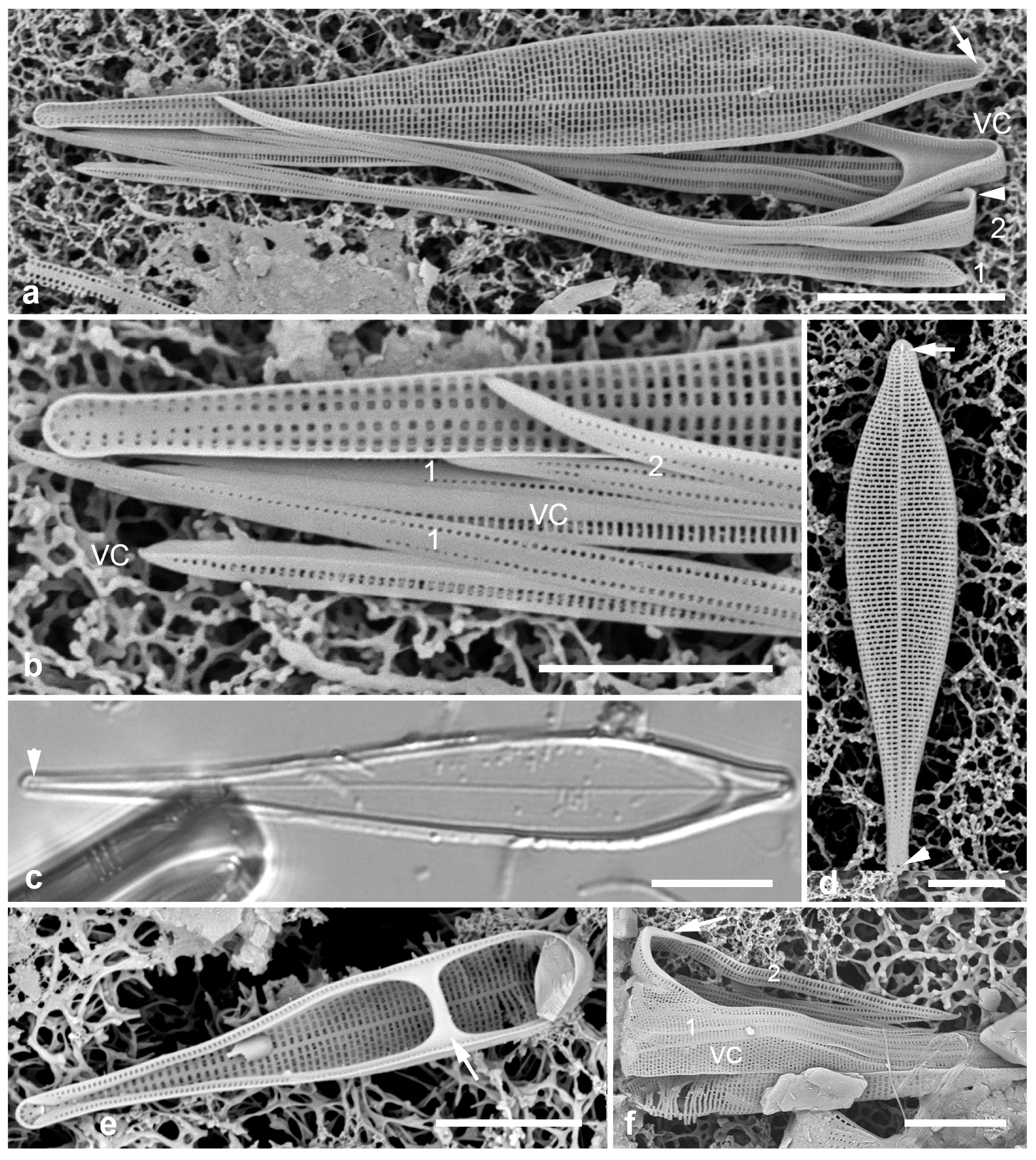
3.68. Licmophora johnwestii Lobban and Emm. S. Santos 2022 [33]—Figure 22e,f
Yap samples: Y26A, C
Dimensions: Length 17–21 μm, width 3.5–4 μm at septal bridge. Stria density different in basal and apical halves of the valve, 32–33 to 43–44 in 10 μm
Comment: The type locality for this distinctive species is Melbourne, Australia, where it was epiphytic on seaweed in an intertidal pool. The Yap samples, from subtidal sediment collections, were likely washed off the reef.
3.69. Licmophora peragallioides Lobban 2013 [112]—Figure 23a
Yap samples: Y41-8
Dimensions (from Guam): Length 68–109 µm, width 14–16 µm, striae 12–13 near base, 14–16 near apex.
Comments: Although recorded only as the valvocopula, it conforms to L. peragallioides.
3.70. Licmophora remulus Grunow 1867 [7]—Figure 23b
Yap samples: Y36-4
Dimensions: Length 84 (100–160) µm, width at apex 8 µm, striae 29–30 in 10 µm
3.71. Licmophora romuli Lobban 2021 [21]—Figure 23c–e
Yap samples: Y41-7
Dimensions: Length 135–182 µm, width 11–12 µm, striae 34 in 10 µm
Diagnostics: With the classic spathulate outline of L. remulus but differing in the dearth of vimines in the apical part, giving it a shredded appearance in SEM; difficult to distinguish from L. remulus in LM.
Licmophora, cont. (a) L. peragallioides, LM, showing apical window in septum (arrow). (b) L. remulus, LM, showing regular areolae in the lamina. (c–e) L. romuli, SEM, valve with details of basal pole and lamina; (d) showing single line of areolae on each side of the sternum on the “stem” with short striae on the basal pole, and 8 slits in the multiscissura; (e) showing the centripetal loss of vimines on the lamina, resulting in shredding of the valve in acid cleaning. (f) L. undulata, LM, showing undulation (arrows), parallel sides of lower stem, and inflated base. Scale bars: (c) = 25 µm, (a,b,e,f) = 10 µm, (d) = 2 µm.
Figure 23.
Licmophora, cont. (a) L. peragallioides, LM, showing apical window in septum (arrow). (b) L. remulus, LM, showing regular areolae in the lamina. (c–e) L. romuli, SEM, valve with details of basal pole and lamina; (d) showing single line of areolae on each side of the sternum on the “stem” with short striae on the basal pole, and 8 slits in the multiscissura; (e) showing the centripetal loss of vimines on the lamina, resulting in shredding of the valve in acid cleaning. (f) L. undulata, LM, showing undulation (arrows), parallel sides of lower stem, and inflated base. Scale bars: (c) = 25 µm, (a,b,e,f) = 10 µm, (d) = 2 µm.
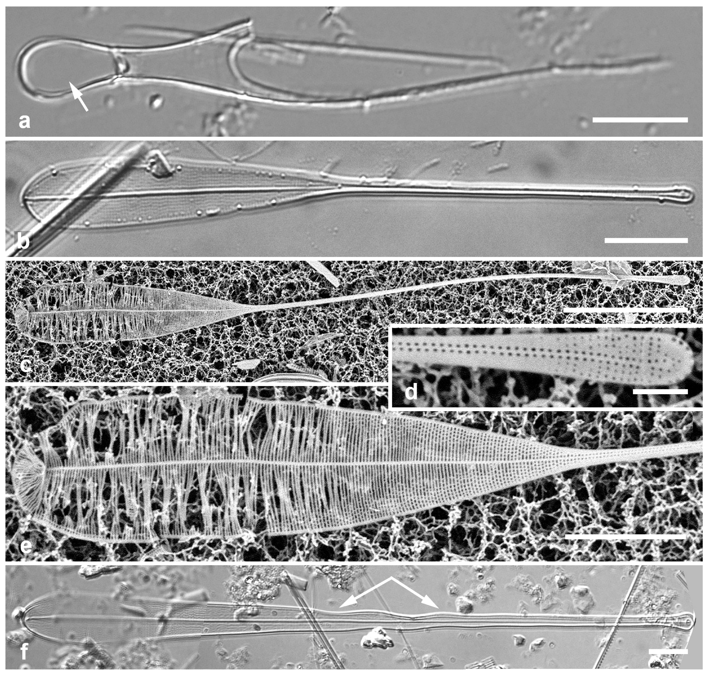
3.72. Licmophora undulata Macatugal, Tharngan & Lobban 2019 [113]—Figure 23f
Yap samples: Y26C
Dimensions: Length 171 µm, width near apex 13 µm, striae 30 in 10 µm
Diagnostics: The long “stem” has a distinct wave in it.
3.73. Podocystis adriatica (Kützing) Ralfs in Prichard 1861 [50]—Figure 24a,b
Yap samples: Y37-7, Y37-8, Y41-8
Dimensions: Length 38–64 µm, width 18–23 µm
Comment: Podocystis spp. attach to seaweeds by mucilage pads.
(a,b) Podocystis adriatica. (a) SEM, external, showing bi- to multiseriate striae. (b) LM showing costae between the striae. (c) Podocystis spathulata SEM, internal, showing absence of costae; this valve has apical and basal rimoportulae (arrowheads), the other valve would have only apical. There is also a characteristic pore near the sternum (arrow). (d,e) Lioloma delicatulum, SEM external valve faces, showing basal pole with rimoportula (arrowhead) and part of the middle of a very long valve. (f–h) Lioloma elongatum, SEM external valve faces, showing basal pole with rimoportula (arrowhead) and portion of the middle with a “bubble-shaped structure” (arrow). Scale bars: (a–c) = 10 µm, (d–f,h) = 5 µm, (g) = 2 µm.
Figure 24.
(a,b) Podocystis adriatica. (a) SEM, external, showing bi- to multiseriate striae. (b) LM showing costae between the striae. (c) Podocystis spathulata SEM, internal, showing absence of costae; this valve has apical and basal rimoportulae (arrowheads), the other valve would have only apical. There is also a characteristic pore near the sternum (arrow). (d,e) Lioloma delicatulum, SEM external valve faces, showing basal pole with rimoportula (arrowhead) and part of the middle of a very long valve. (f–h) Lioloma elongatum, SEM external valve faces, showing basal pole with rimoportula (arrowhead) and portion of the middle with a “bubble-shaped structure” (arrow). Scale bars: (a–c) = 10 µm, (d–f,h) = 5 µm, (g) = 2 µm.
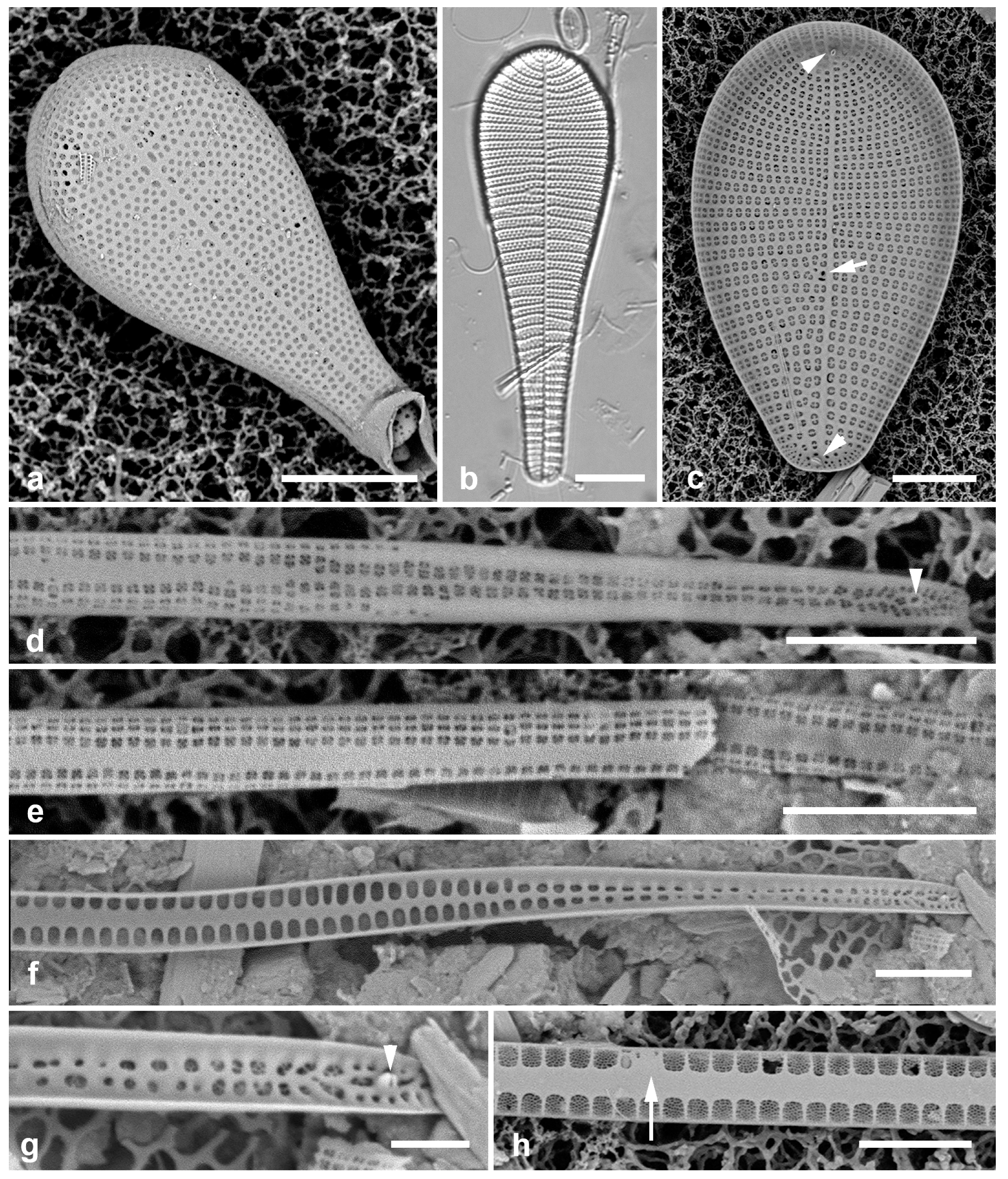
3.74. Podocystis spathulata (Shadbolt) Van Heurck 1896 [114]—Figure 24c
Yap samples: Y25H-2, Y36-2, Y37-8, Y45-2
Thalassionemataceae Round
3.75. Lioloma delicatulum (Cupp) Hasle 1996 in [115]—Figure 24d,e
Yap samples: Y26B
Dimensions: Valves, 3 µm wide away from the apex, are said to be 900 µm long, but we did not find any complete valves.
Diagnostics: Very long slender valves without marginal spines, with several areolae in short striae on the mantle and valve face.
Comments: Planktonic. We observed only the basal pole. Lioloma differs from Thalassiothrix in lacking marginal spines and the genus name reflects this (“smooth margin”).
3.76. Lioloma elongatum (Grunow) Hasle 1996 in [115]—Figure 24f–h
Yap samples: Y26B
Dimensions: Valves, 3 µm wide away from the apex, are said to be 900–2000 µm long, but no complete valves found.
Comments: We observed only the basal pole. This species differs from L. delicatulum in the number of areolae in a stria. Planktonic.
3.77. Thalassionema baculum Lobban 2021 [21]—Figure 25b,c
Yap samples: Y26B, Y26C
Dimensions: Length 19 µm, width 2.5 µm, striae 10 in 10 µm
Diagnostics: Small, baton-like frustule, with single line of areolae along the valve margin, simple bar across each areola.
Comments: The only species in this genus commonly observed in the region, and probably the only benthic species.
3.78. Thalassionema synedriforme (Greville) Hasle 1999 [117]—Figure 25a
Yap samples: Y26C
Comments: Planktonic. Type locality: Hong Kong harbor.
3.79. Thalassiothrix gibberula Hasle 1996 in [115]—Figure 25d–g
Yap samples: Y26B
Dimensions: Length > 830 µm, width 1.7–2.7 µm, marginal spines ca. 30 in 100 µm
Diagnostics: Very long slender valves with dense marginal spines pointed toward the head pole, wide sternum with a longitudinal rib plus spines separating the areolae along the valve margin.
(a) Thalassionema synedriforme, SEM portion with apical spine. (b,c) Thalassionema baculum. (b). Frustule in LM. (c) Frustules in girdle view showing simple bars across areolae. (d–g) Thalassiothrix gibberula. (d) Portion of valve in LM showing prominent spines. (e) Basal pole with characteristic spines, SEM. (f) Portions of valves showing interior and exterior. (g) Fragments of valve in external view. Scale bars: (b,d,f) = 10 µm, (a,c,e,g) = 5 µm.
Figure 25.
(a) Thalassionema synedriforme, SEM portion with apical spine. (b,c) Thalassionema baculum. (b). Frustule in LM. (c) Frustules in girdle view showing simple bars across areolae. (d–g) Thalassiothrix gibberula. (d) Portion of valve in LM showing prominent spines. (e) Basal pole with characteristic spines, SEM. (f) Portions of valves showing interior and exterior. (g) Fragments of valve in external view. Scale bars: (b,d,f) = 10 µm, (a,c,e,g) = 5 µm.

3.80. Gato hyalinus Lobban & Navarro 2013 [118]—Figure 26a,b
Yap samples: Y37-7, Y37-8, Y36-1
Dimensions: Length 43–52 µm, width 16–18 µm, striae 60–70 in 10 µm (not resolved in LM)
(a,b) Gato hyalinus, LM showing hyaline valve face with apical rimoportula (arrow), and SEM external valve face showing very fine striae and opening of apical rimoportula (arrow). (c–g) Glyphodesmis acus. (c–e) Yap specimens in LM (same scale). (f) Guam specimen (GU52P-7) in LM. (g) Yap specimen in SEM, internal face and narrow copulae. Scale bars: (a–f) = 10 µm, (g) = 5 µm.
Figure 26.
(a,b) Gato hyalinus, LM showing hyaline valve face with apical rimoportula (arrow), and SEM external valve face showing very fine striae and opening of apical rimoportula (arrow). (c–g) Glyphodesmis acus. (c–e) Yap specimens in LM (same scale). (f) Guam specimen (GU52P-7) in LM. (g) Yap specimen in SEM, internal face and narrow copulae. Scale bars: (a–f) = 10 µm, (g) = 5 µm.
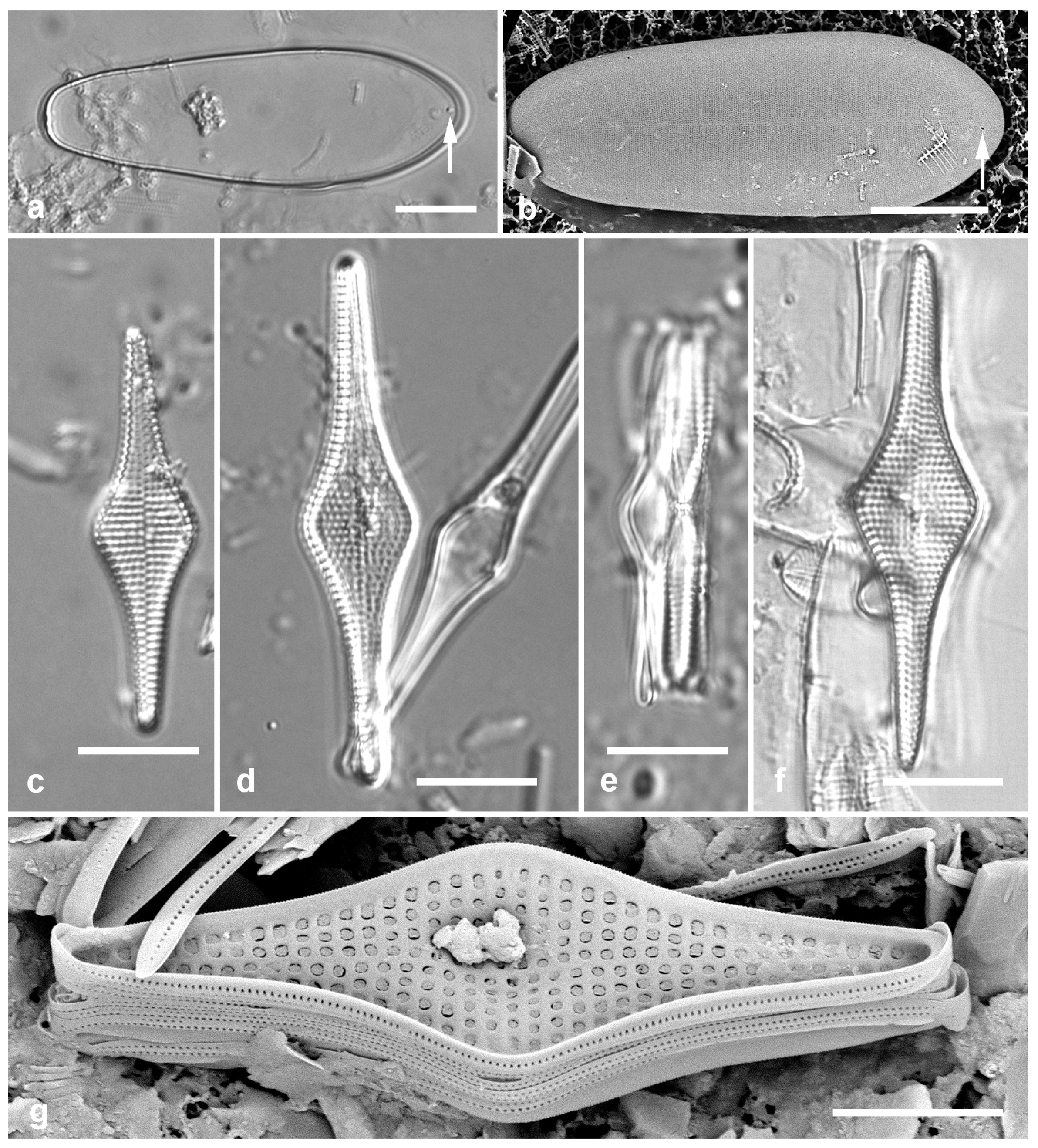
3.81. Glyphodesmis acus A. Mann 1925 [9]—Figure 26c–g and Figure 27
Yap samples: Y26C
Dimensions: Length 32–43 µm, width 10–11 µm; striae 15 in 10 µm.
Glyphodesmis acus, cont., Guam specimens (GU52P-7), SEM. (a) Valve, internal view. (b) Frustule, oblique internal view, showing interlocking spines between two valves, multiple copulae (some hyaline, others perforate), and elevated center. (c) Detail of same specimen to show apical pore fields (arrows). (d) Portion of different frustule in girdle view near center, showing copulae and overlapping spines. Scale bars: (a,b) = 10 µm, (d) = 5 µm, (c) = 2 µm.
Figure 27.
Glyphodesmis acus, cont., Guam specimens (GU52P-7), SEM. (a) Valve, internal view. (b) Frustule, oblique internal view, showing interlocking spines between two valves, multiple copulae (some hyaline, others perforate), and elevated center. (c) Detail of same specimen to show apical pore fields (arrows). (d) Portion of different frustule in girdle view near center, showing copulae and overlapping spines. Scale bars: (a,b) = 10 µm, (d) = 5 µm, (c) = 2 µm.
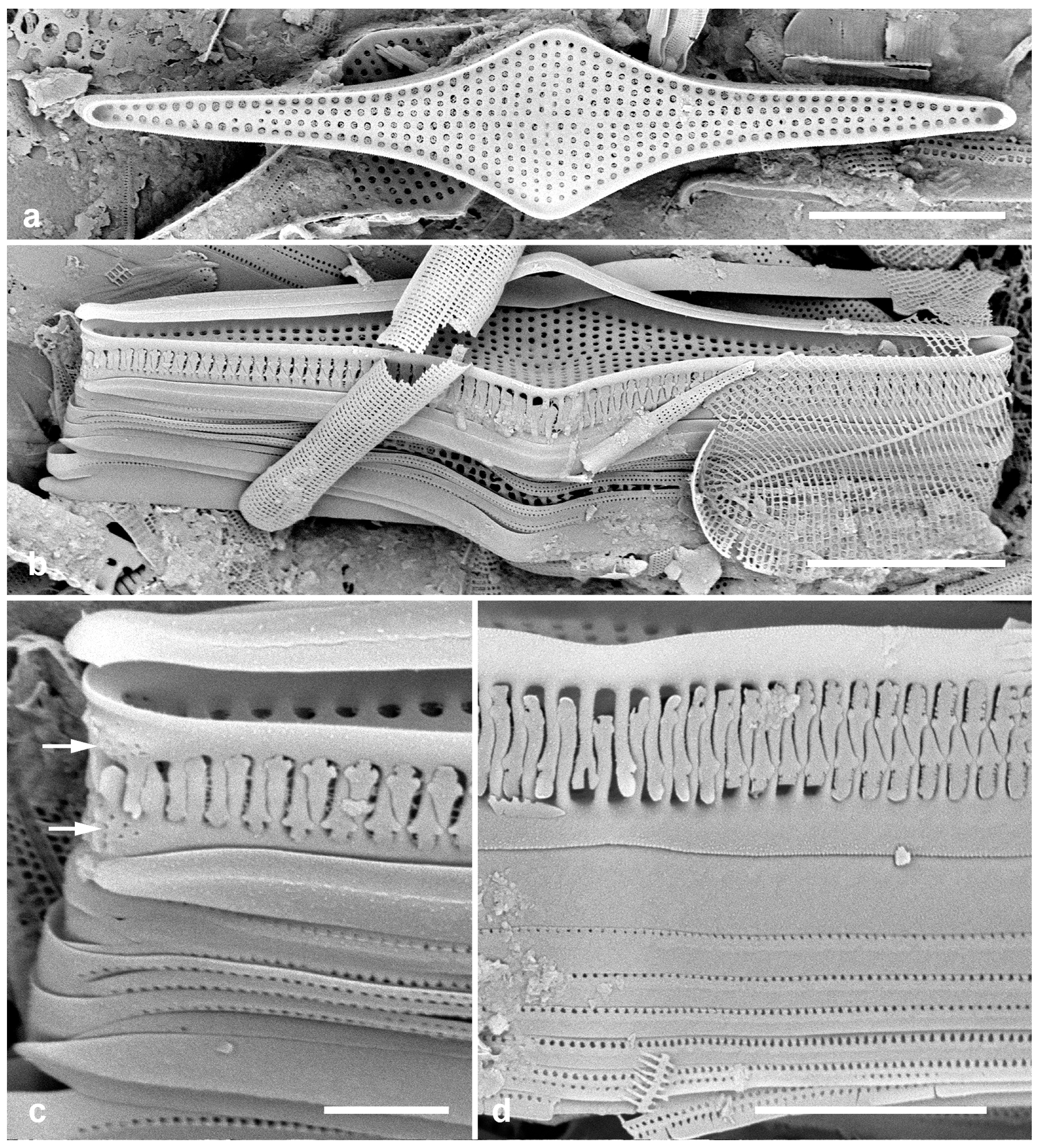
Diagnostics: Valves rhomboidal to linear–lanceolate with strongly inflated center, center of the valve elevated but without distinctive structure. Sternum absent. SEM shows small pore fields on valve apex and mantle but no ocellus. Interlocking spines at end of each stria, no areolae on mantle; spines resemble those of Hendeyella lineata. Copulae of various widths but generally narrow, 0–2 rows of pores on each. Apparently no rimoportulae. External valve face not seen.
3.82. Colliculoamphora gabgabensis Lobban 2015 [13]—Figure 28a,b
Yap samples: Y36-5, Y37-8, Y41-8
3.83. Lyrella clavata (Gregory) D.G.Mann 1990 in [52]—Figure 28c
Yap samples: Y26C
Dimensions: Length 62–77 µm, width 38–45 µm, striae 14–15 in 10 µm
(a,b) Colliculoamphora gabgabensis, frustule in SEM and valve in LM. (c) Lyrella clavata, LM. (d,e) Lyrella cf. rudiformis, SEM, valve exterior and detail of areolae. (f) Lyrella lyra, L.M. (g) Lyrella clavata valve exterior, SEM. Scale bars: (a,c,f,g) = 10 µm, (d) = 5 µm, (b) = 2 µm, (e) = 1 µm.
Figure 28.
(a,b) Colliculoamphora gabgabensis, frustule in SEM and valve in LM. (c) Lyrella clavata, LM. (d,e) Lyrella cf. rudiformis, SEM, valve exterior and detail of areolae. (f) Lyrella lyra, L.M. (g) Lyrella clavata valve exterior, SEM. Scale bars: (a,c,f,g) = 10 µm, (d) = 5 µm, (b) = 2 µm, (e) = 1 µm.
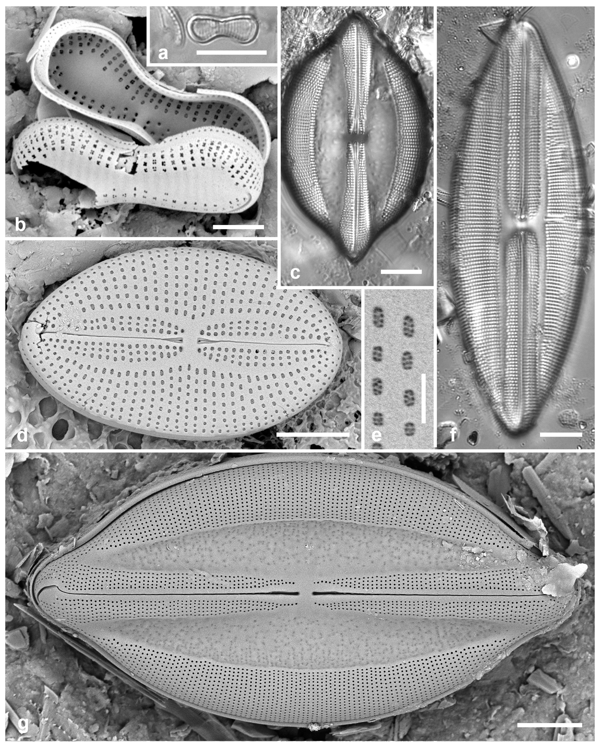
3.84. Lyrella lyra (Ehrenberg) Karayeva 1978 [126]—Figure 28f
Yap samples: Y25H-1, Y25H-2, Y36-1
Dimensions: Length 98 µm, width 33 µm, striae 13 in 10 µm
Diagnostics: Distinguished from other Lyrella spp. by the narrow, straight lateral sterna.
3.85. Lyrella cf. rudiformis (Hustedt) Gusliakov & Karaeva 1992 in [127]—Figure 28d,e
Samples: Y26C
Dimensions: Length 22 µm, width 13 µm, striae 16 in 10 µm
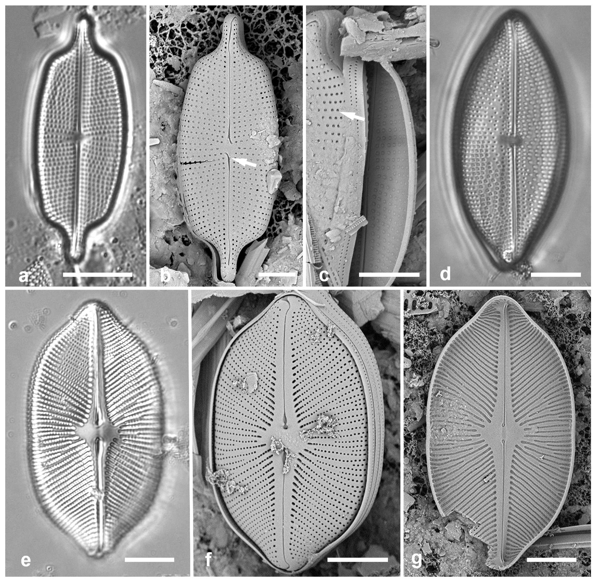
3.86. Moreneis cf. hexagona J.Park, Koh & Witkowski 2012 in [129]—Figure 29a–c
Yap samples: Y26C, Y42-1
Dimensions: Length 35 µm, width 13 µm, striae 16 in 10 µm
3.87. Petroneis granulata (Bailey) D.G. Mann 1990 in [52]—Figure 29d
Yap samples: Y26B, Y37-7
Dimensions: Length 54 µm, width 25 µm, striae 11 in 10 µm
3.88. Petroneis humerosa (Brébisson ex W. Smith) Stickle & D.G. Mann 1990 in [52]—Figure 29e–g
Yap samples: Y16B, Y25H-2
Dimensions: Length 46–58 µm, width 28–30 µm, striae 10 in 10 µm
(a–c) Moreneis cf. hexagona. (a) Valve in LM. (b) Valve in SEM, external view showing characteristic central raphe endings (arrow). (c) Broken frustule in SEM showing interior covered foramina. Also note the single bar extending into each areola (arrow). (d) Petroneis granulata, LM. (e–g) Petroneis humerosa. (e) Valve in LM. (f) Frustule in SEM, external also showing copulae. (g) Valve internal surface in SEM. Scale bars: (a,d–g) = 10 µm, (b,c) = 5 µm.
Figure 29.
(a–c) Moreneis cf. hexagona. (a) Valve in LM. (b) Valve in SEM, external view showing characteristic central raphe endings (arrow). (c) Broken frustule in SEM showing interior covered foramina. Also note the single bar extending into each areola (arrow). (d) Petroneis granulata, LM. (e–g) Petroneis humerosa. (e) Valve in LM. (f) Frustule in SEM, external also showing copulae. (g) Valve internal surface in SEM. Scale bars: (a,d–g) = 10 µm, (b,c) = 5 µm.
Mastogloiaceae Mereschkowsky
3.89. Mastogloiopsis biseriata Lobban & Navarro 2012 [131]—Figure 30a
Yap samples: Y26C
Dimensions: Length 25 µm, striae 33 in 10 µm.
(a) Mastogloiopsis biseriata, frustule in girdle view, SEM. (b,c) Tetramphora decussata internal view in SEM, valve in LM. (d) Tetramphora intermedia, LM. (e,f) Dictyoneis cf. marginata, frustules in girdle and oblique views, LM. Scale bars: (b–d) = 25 µm, (e,f) = 10 µm, (a) = 5 µm.
Figure 30.
(a) Mastogloiopsis biseriata, frustule in girdle view, SEM. (b,c) Tetramphora decussata internal view in SEM, valve in LM. (d) Tetramphora intermedia, LM. (e,f) Dictyoneis cf. marginata, frustules in girdle and oblique views, LM. Scale bars: (b–d) = 25 µm, (e,f) = 10 µm, (a) = 5 µm.
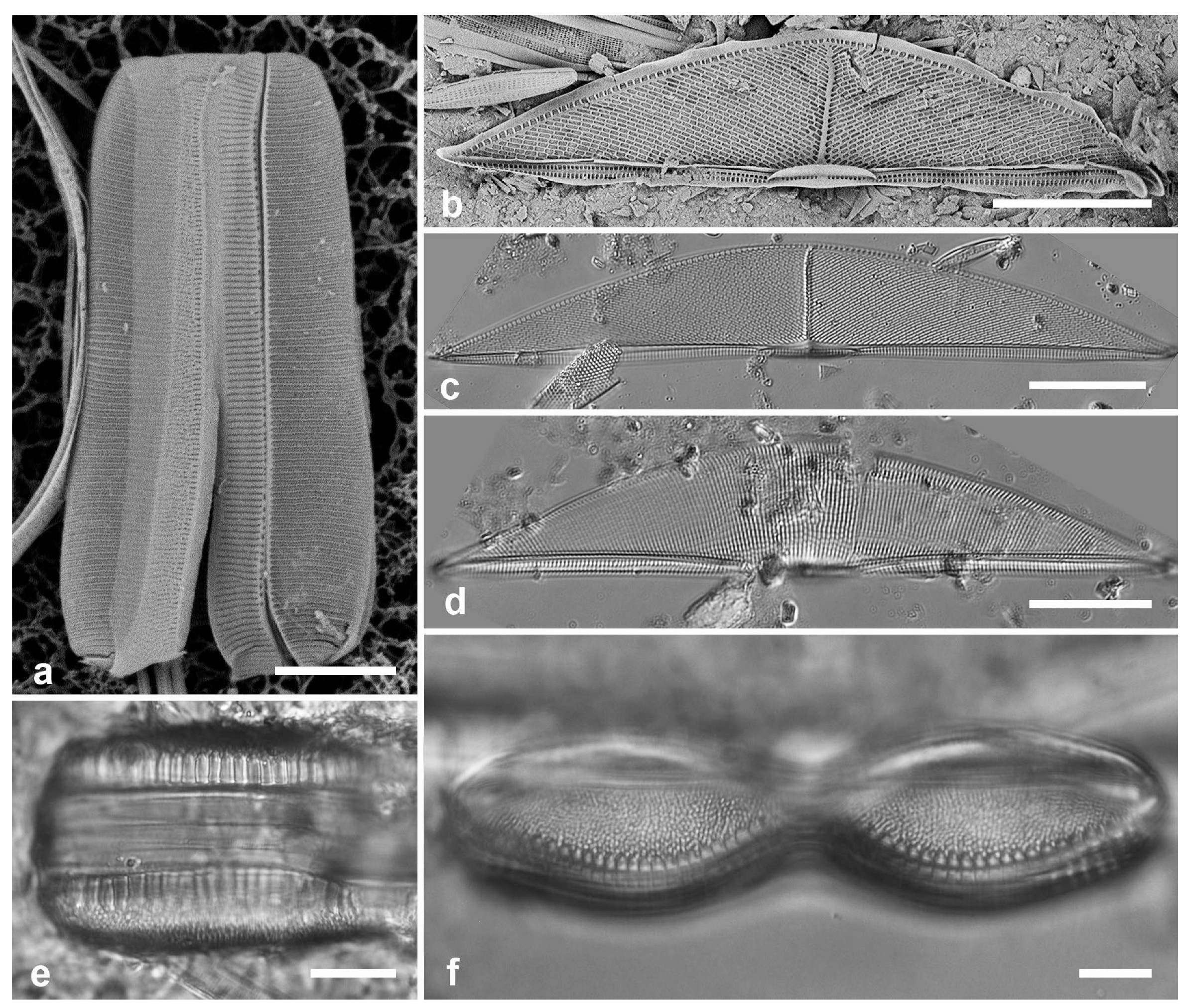
3.90. Tetramphora decussata (Grunow) Stepanek & Kociolek 2016 [132]—Figure 30b,c
Yap samples: Y25H-2, Y26B, Y26C
Dimensions: Length 115–144 µm, width 22–25 µm, dorsal striae 19 in 10 µm
3.91. Tetramphora intermedia (Cleve) Stepanek & Kociolek 2016 [132]—Figure 30d
Yap samples: Y25H-2, Y42-1
Dictyoneidaceae D.G. Mann
3.92. Dictyoneis cf. marginata (F.W.Lewis) Cleve 1890 [133]—Figure 30e,f
Yap samples: Y26C
Dimensions: Length 82–103 µm
Diagnostics: Panduriform frustules with coarse apparent poration caused by pseudoloculate structure; large openings along the margin suggestive of Mastogloia partecta.
Rhoicospheniaceae Chen & Zhu
3.93. Gomphonemopsis littoralis (Hendey) Medlin 1986 in [134]—Figure 31a
Yap samples: Y41-7
Dimensions: Length 12 µm, width 2 µm, striae 20–21 in 10 µm.
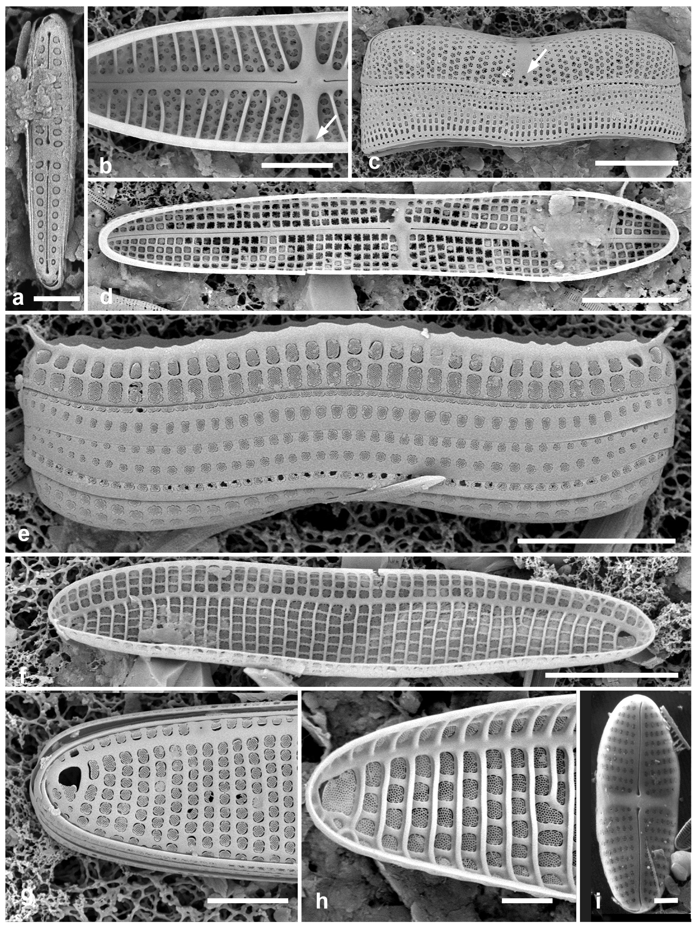
3.94. Achnanthes armillaris (O.F.Müller) Guiry 2019 [138]—Figure 31b,c
Synonym Achnanthes longipes C.Agardh, nom. illeg.
Yap samples: Y26C
Dimensions: Length 10–40 µm, width 8–10 µm, striae 12–13 in 10 µm (costae 6–8 in 10 µm)
(a) Gomphonemopsis littoralis, SEM. (b,c) Achnanthes armillaris. (b) Internal view of raphe valve showing costae and stauros forked under mantle (arrow). (c) Girdle view of raphe valve and cingulum showing mantle areolae in stauros fork (arrow). (d,e) Achnanthes cf. brevipes. (d) Internal view of raphe valve showing flat stauros and lack of costae. (e) Frustule in girdle view showing mantle rim with apical spines of sternum valve. (f–h) Achnanthes kuwaitensis. (f) Internal view of sternum valve. (g) External detail of sternum valve showing apical obiculus (cribrum mostly missing). (h) Internal detail of sternum valve apex with intact cribrum in obiculus. (i) Achnanthes parvula, Yap voucher, SEM (courtesy of Nelson Navarro). Scale bars: (c–f) = 10 µm, (b,g) = 5 µm, (a,h,i) = 2 µm.
Figure 31.
(a) Gomphonemopsis littoralis, SEM. (b,c) Achnanthes armillaris. (b) Internal view of raphe valve showing costae and stauros forked under mantle (arrow). (c) Girdle view of raphe valve and cingulum showing mantle areolae in stauros fork (arrow). (d,e) Achnanthes cf. brevipes. (d) Internal view of raphe valve showing flat stauros and lack of costae. (e) Frustule in girdle view showing mantle rim with apical spines of sternum valve. (f–h) Achnanthes kuwaitensis. (f) Internal view of sternum valve. (g) External detail of sternum valve showing apical obiculus (cribrum mostly missing). (h) Internal detail of sternum valve apex with intact cribrum in obiculus. (i) Achnanthes parvula, Yap voucher, SEM (courtesy of Nelson Navarro). Scale bars: (c–f) = 10 µm, (b,g) = 5 µm, (a,h,i) = 2 µm.
Achnanthes cf. brevipes, Pohnpei population (PN1-1). (a) General view of population on filamentous seaweed, frustules attached by raphe valves. (b) Frustule in girdle view showing rim with spines on sternum valve. Scale bars: (a) = 50 µm, (b) = 10 µm.
Figure 32.
Achnanthes cf. brevipes, Pohnpei population (PN1-1). (a) General view of population on filamentous seaweed, frustules attached by raphe valves. (b) Frustule in girdle view showing rim with spines on sternum valve. Scale bars: (a) = 50 µm, (b) = 10 µm.
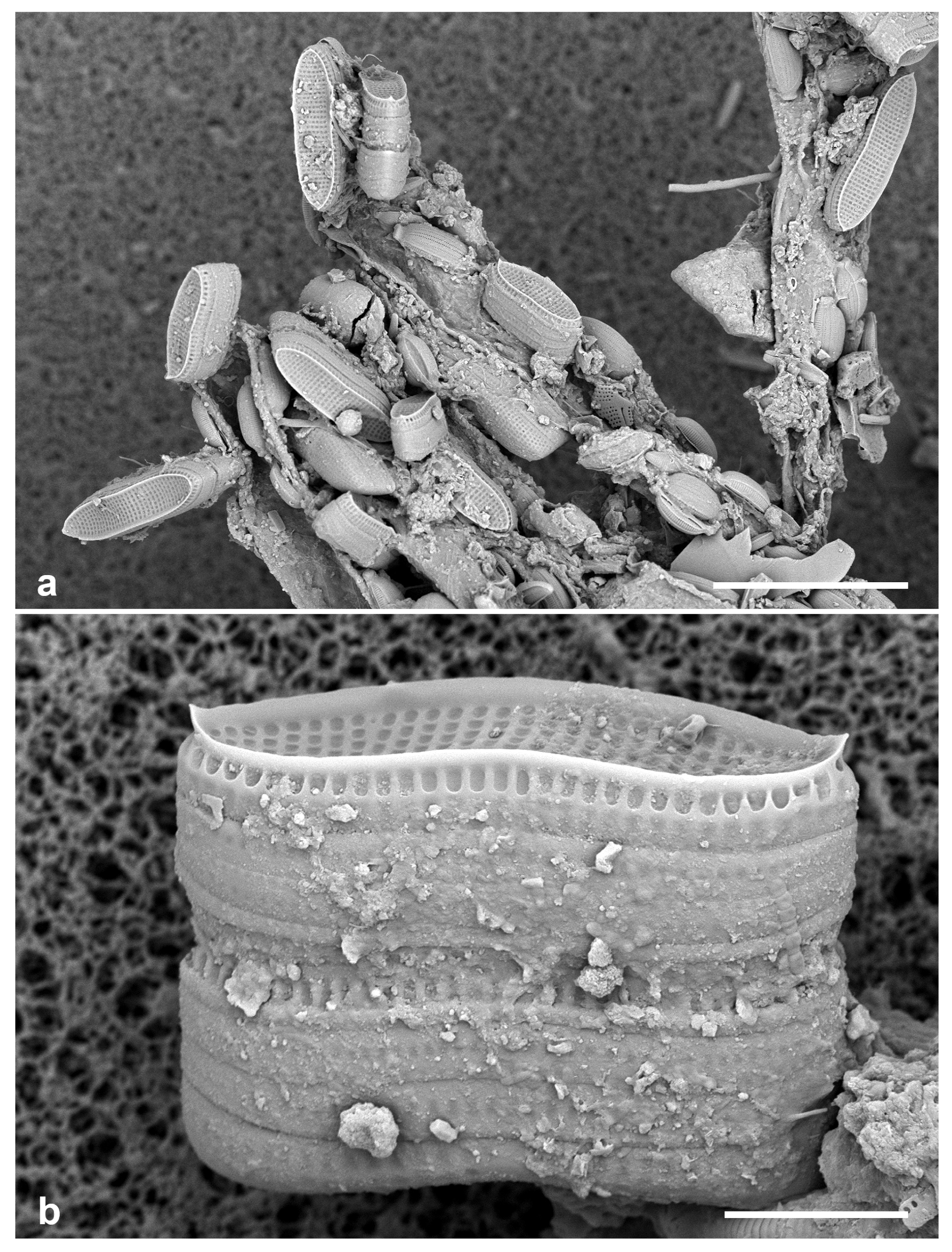
3.95. Achnanthes cf. brevipes C.A. Agardh 1824 [140]—Figure 31d,e and Figure 32
Yap samples: Y16B, Y16N, Y26C
Dimensions: Length 36–77 µm, width 9–14 µm, striae 11 in 10 µm
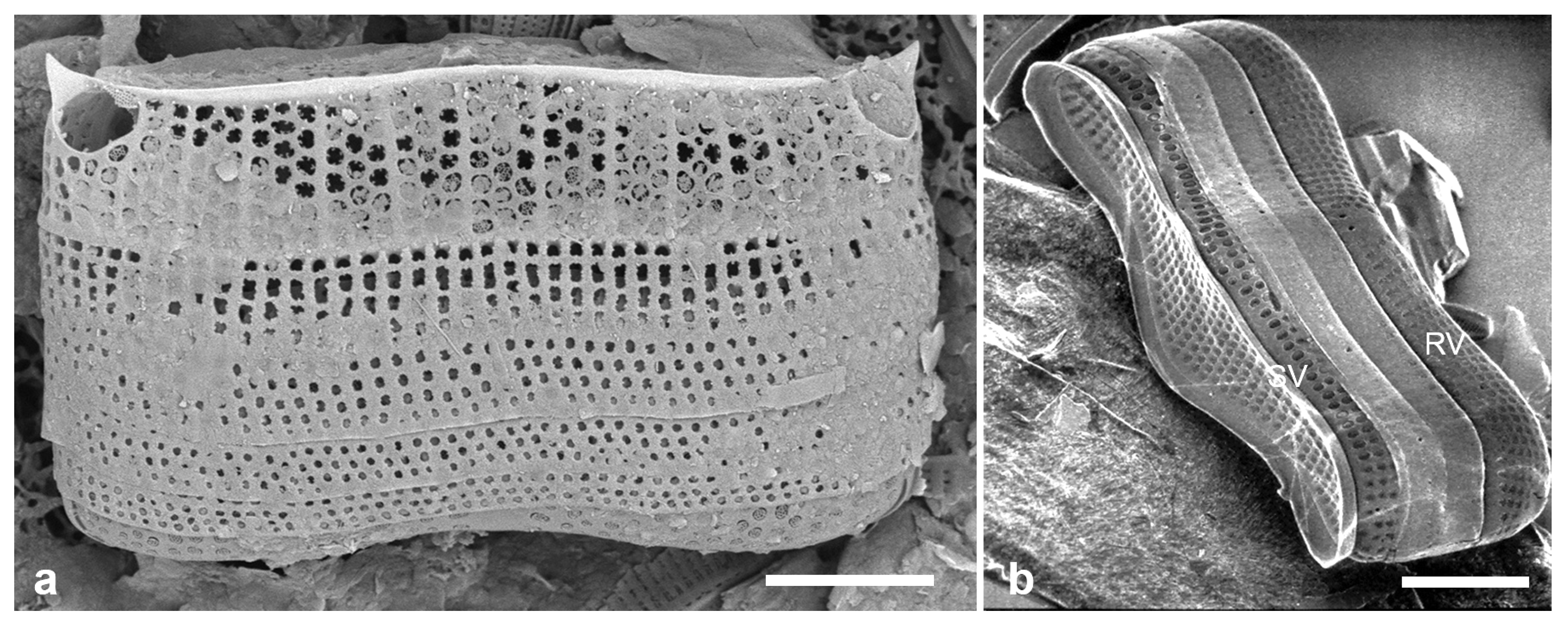
(a) Achnanthes (undescribed sp.?) frustule from Yap in girdle view, showing spines and large areolae on mantle (cribrum lost on left, obscured on right). (b) A. inflata (freshwater) showing difference in copulae as well as the inflated center; SV = sternum valve, RV = raphe valve (Yap voucher, courtesy of N. Navarro). Scale bars: (b) = 10 µm, (a) = 5 µm.
Figure 33.
(a) Achnanthes (undescribed sp.?) frustule from Yap in girdle view, showing spines and large areolae on mantle (cribrum lost on left, obscured on right). (b) A. inflata (freshwater) showing difference in copulae as well as the inflated center; SV = sternum valve, RV = raphe valve (Yap voucher, courtesy of N. Navarro). Scale bars: (b) = 10 µm, (a) = 5 µm.
3.96. Achnanthes kuwaitensis Hendey 1958 [142]—Figure 31f–h
Dimensions: Length 43–49 µm, width 9–10 µm, striae 9–10 in 10 µm.
Diagnostics: The linear valve outline with the large obiculi at each end of the sternum valve, the sternum on the valve face–mantle boundary, and the internal costae distinguish this species from A. brevipes and from other species with obiculi.
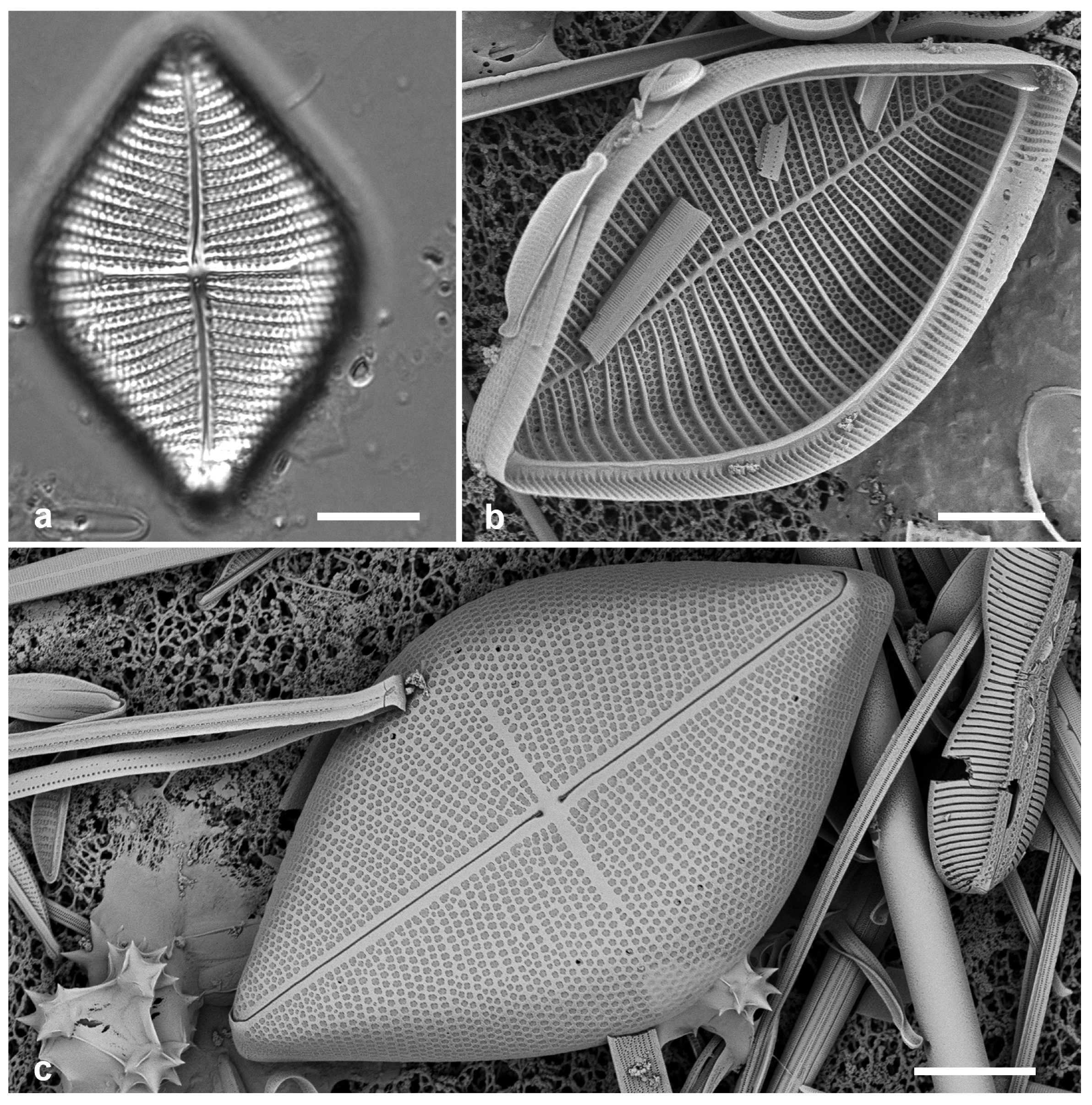
3.97. Achnanthes grunowii Toyoda & D.M. Williams in [143]—Figure 34
Synonym: Achnanthes javanica var. rhombica Grunow
Yap samples: Y26C
Dimensions: Length 49 µm, width, 33 µm, striae biseriate, 7 in 10 µm.
3.98. Achnanthes orientalis F.Meister 1937 [48]—Figure 35
non Achnanthes orientalis Petit 1904, nec Achnanthes orientalis Hustedt 1933
Yap samples: Y26C, Y41-8
Dimensions: Length 36–66 µm, width 8–12.5 µm; striae 13–15 in 10 µm
Diagnostics: Lanceolate–rhombic valves, slightly flexed toward poles, RV convex, SV concave; apices turned slightly in opposite directions, sometimes rostrate; raphe path sinuous; striae biseriate as seen in SEM. Stria densities similar on sternum and raphe valves, but striae shorter on SV, leaving a broad lanceolate hyaline area in the middle of the valve.
3.99. Achnanthes parvula Kützing 1844 [37]—Figure 31i
Achnanthes grunowii. (a) Raphe valve in LM. (b,c) Valves in SEM (Palau specimens, PW2009-22). (b) Sternum valve interior with valvocopula. (c) Raphe valve exterior (with Diploneis crispa). Scale bars: 10 µm.
Figure 34.
Achnanthes grunowii. (a) Raphe valve in LM. (b,c) Valves in SEM (Palau specimens, PW2009-22). (b) Sternum valve interior with valvocopula. (c) Raphe valve exterior (with Diploneis crispa). Scale bars: 10 µm.
Achnanthes orientalis. (a) Exterior views of rostrate raphe and sternum valves (Palau, PW46). (b–d) Yap specimens with more typical apices in LM and SEM. (e) Part of frustule in oblique view showing apical curvature of sternum valve (Chuuk, TK1A). Scale bars: (a–d) = 10 µm, (e) = 5 µm.
Figure 35.
Achnanthes orientalis. (a) Exterior views of rostrate raphe and sternum valves (Palau, PW46). (b–d) Yap specimens with more typical apices in LM and SEM. (e) Part of frustule in oblique view showing apical curvature of sternum valve (Chuuk, TK1A). Scale bars: (a–d) = 10 µm, (e) = 5 µm.

Achnanthidiaceae D.G. Mann
3.100. Planothidium delicatulum (Kützing) Round & Bukhtiyarova 1996 [145]—Figure 36a–c
Yap samples: Y16B
Dimensions: Length 10 µm, width 5 µm, striae ca. 12 in 10 µm.
Diagnostics: Small rostrate valves with radiate multiseriate striae, no central break in striae.
(a–c) Planothidium delicatulum, SEM: raphe valve (RV) interior and exterior, sternum valve interior. (d–f) Anorthoneis sp. (d) Sternum valve (SV) in LM. (e,f) SV in SEM, with and without papillae. (g) Cocconeis convexa, RV and SV in LM. Scale bars: (d,g) = 10 µm, (e,f) = 5 µm, (a–c) = 2 µm.
Figure 36.
(a–c) Planothidium delicatulum, SEM: raphe valve (RV) interior and exterior, sternum valve interior. (d–f) Anorthoneis sp. (d) Sternum valve (SV) in LM. (e,f) SV in SEM, with and without papillae. (g) Cocconeis convexa, RV and SV in LM. Scale bars: (d,g) = 10 µm, (e,f) = 5 µm, (a–c) = 2 µm.
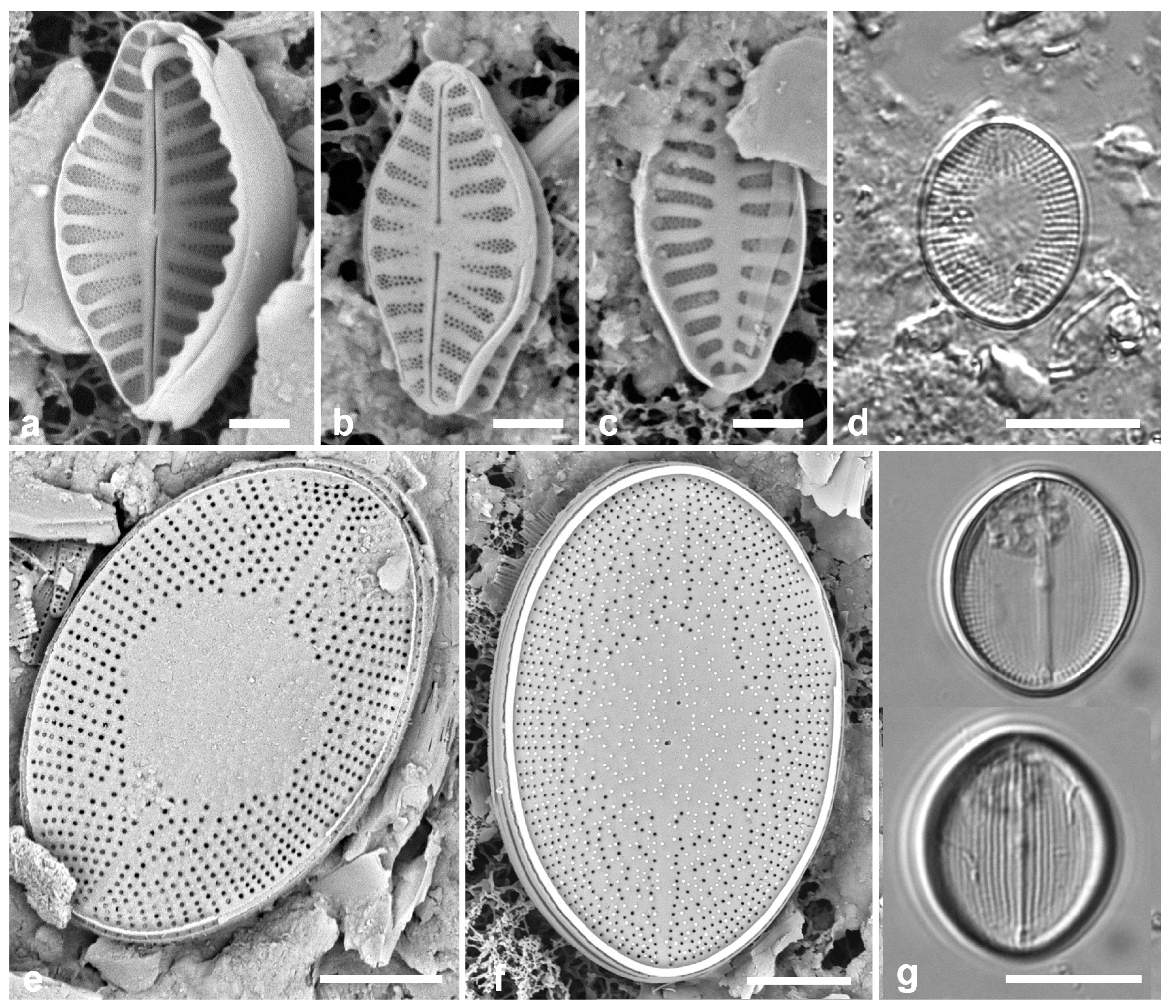
3.101. Anorthoneis sp.—Figure 36d–f
Yap samples: Y16B, Y26C, Y34E
Dimensions: Length 17–25 µm, width 12–16 µm; radiate striae 18–19 in 10 µm (SV).
Diagnostics: Oval valves; sternum valve with oval hyaline zone in the middle.
3.102. Cocconeis convexa Giffen 1967 [149]—Figure 36g and Figure 37a,b
Yap samples: Y25H-1, Y25H-2, Y26C, Y37-8, Y37-7, Y36-1
Dimensions: Length 17–19 µm, width 13 µm; striae on SV 40 in 10 µm, on RV 21 in 10 µm
3.103. Cocconeis coronatoides Riaux-Gobin & Romero 2011 in [150]—Figure 37c
Yap samples: Y25H-1, Y25H-2, Y26C, Y34B, Y37-8, Y37-7, Y36-1, Y D-2
Dimensions: Length 29 µm, width 19 µm, striae 15 in 10 µm
Diagnostics: Distinguished by the papillae and the submarginal costa on outer SV surface.
3.104. Cocconeis dirupta Gregory 1857 [151]—Figure 37d,e
Yap samples: Y25H-1, Y37-8, Y41-7
Dimensions: Length 11 µm, width 8 µm, striae 24 in 10 µm (SV), not resolved in LM on RV
3.105. Cocconeis heteroidea Hantzsch 1863 [153]—Figure 37f
(a,b) Cocconeis convexa, cont. (a) Sternum valve, exterior, SEM. (b) Raphe valve exterior (Majuro), showing convexity (whole frustule is curved to fit on algal filaments, so this lower surface is concave). (c) Cocconeis coronatoides, sternum valve, SEM. (d,e) Cocconeis dirupta, LM of RV and SV, respectively. (f) Cocconeis heteroidea frustule at two focal planes in LM, showing RV and SV. (g,h) Berkeleya rutilans. Scale bars: (f) = 10 µm, (a–e,g,h) = 5 µm.
Figure 37.
(a,b) Cocconeis convexa, cont. (a) Sternum valve, exterior, SEM. (b) Raphe valve exterior (Majuro), showing convexity (whole frustule is curved to fit on algal filaments, so this lower surface is concave). (c) Cocconeis coronatoides, sternum valve, SEM. (d,e) Cocconeis dirupta, LM of RV and SV, respectively. (f) Cocconeis heteroidea frustule at two focal planes in LM, showing RV and SV. (g,h) Berkeleya rutilans. Scale bars: (f) = 10 µm, (a–e,g,h) = 5 µm.

Yap samples: Y25H-1, Y25H-2, Y37-8, Y41-7, Y41-8
3.106. Berkeleya rutilans (Trentepohl ex Roth) Grunow 1880 [154]—Figure 37g,h
Yap samples: Y26A
Dimensions: Valves 18 µm long, 3.5 µm wide, striae 32 in 10 µm near along the raphe branches, coarser in middle.
3.107. Climaconeis lorenzii Grunow 1862 [158]
Dimensions: Length 140–146 µm, width 7 µm, striae 15 in 10 µm
Diagnostics: Straight cells, central raphe endings deflected, no stauros; craticular bars present on valvocopula.
3.108. Climaconeis minaegensis Lobban 2021 [21]
Yap samples: Y18E
Dimensions: Length 228–247 µm, width 5.0 µm except 8.3 µm at center and 7.6 µm at apex. Striae 19 in 10 µm
Diagnostics: Valves long, linear but slightly bent along the apical plane, without craticular bars or stauros, silica ribs bordering the raphe, quadrate to transapically elongate areolae.
3.109. Climaconeis tarangensis Lobban 2021 [21]
Dimensions: Length 121–124 µm, width 4.5 µm; stria density 2021 in 10 µm
Diagnostics: Curved species without craticular bars or stauros, differing from Climaconeis riddleae A.K.S.K. Prasad, in its lower stria density (20 vs. 24–27 in 10 µm), regular apically rectangular areolae, and more strongly arcuate raphe.
Yap samples: Y26C
3.110. Parlibellus biblos (Cleve) E.J.Cox 1988 [159]—Figure 38a,b
Yap samples: Y36-2, Y37-7, Y42-1
Dimensions: Length 28–38 µm, width 4 µm, striae 37 in 10 µm
3.111. Parlibellus hamulifer (Grunow) E.J.Cox 1988 [160]—Figure 38c
Yap samples: Y25H-1, Y26C, Y37-8, Y37-7
Dimensions: Length 47–80 µm, width 8–19 µm, striae 21–22 in 10 µm
Comments: Tube-dwelling species but very mobile and often encountered as solitary cells.
3.112. Parlibellus waabensis Lobban 2021 [21]—Figure 38d and Figure 39
Dimensions: Length 58–64 µm, width 18 µm, striae 22 in 10 µm, coarser in middle
(a,b) Parlibellus biblos, LM and internal SEM. (c,d) Parlibellus hamulifer, LM and internal SEM. (e) Parlibellus waabensis, SEM of Palau specimen, girdle view showing spacing of central striae and larger pores on copulae near mid-cell. Scale bars: (a,c–e) = 10 µm, (b) = 5 µm.
Figure 38.
(a,b) Parlibellus biblos, LM and internal SEM. (c,d) Parlibellus hamulifer, LM and internal SEM. (e) Parlibellus waabensis, SEM of Palau specimen, girdle view showing spacing of central striae and larger pores on copulae near mid-cell. Scale bars: (a,c–e) = 10 µm, (b) = 5 µm.

Scoliotropidaceae Mereschkowsky
3.113. Progonoia diatreta Lobban 2015 [161]—Figure 40a
Yap samples: Y26B, Y26C
Dimensions: Length 52 µm, width 20 µm, striae 7.5 in 10 µm
Comment: The two Progonoia spp. are associated with biofilm on sediments.
3.114. Progonoia intercedens (A. Schmidt) Lobban 2015 [161]—Figure 40b,c
Yap samples: Y26C, Y41-8
3.115. Caloneis egena (A. Schmidt) Cleve 1894 [162]—Figure 41a
Yap samples: Y37-8
Dimensions: Length 27 µm, width 5 µm, striae ca 35 in 10 µm
3.116. Caloneis ophiocephala (Cleve & Grove) Cleve 1894 [162]—Figure 41b
Yap samples: Y26C
Dimensions: Length 68 µm, width 14 µm, striae 18 in 10 µm
Comment: New record for Micronesia.
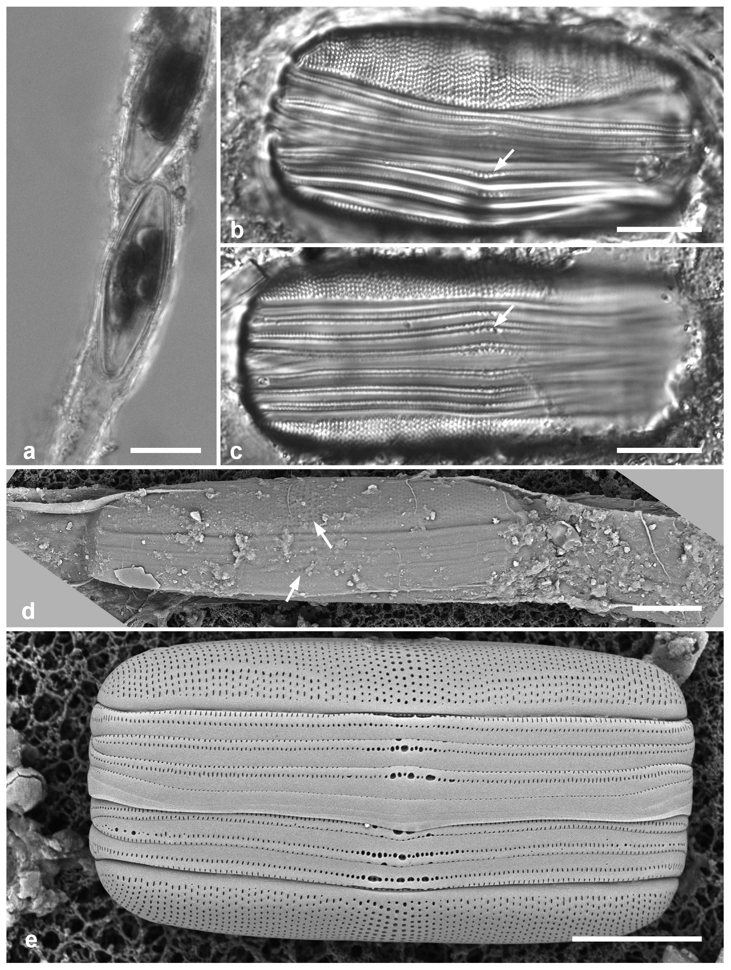
Parlibellus waabensis from Pohnpei (PN1-1) (a,c–e) and Palau (PW(2021)4-7) (b). (a) Preserved cells in mucilage tube, LM. (b,c) Frustules in girdle view showing the distinctive large pores in the girdle bands (arrows), LM. (d) SEM of frustule in mucilage tube, the distinctive copula pores and wider striae spacing on center of cell visible through the dried mucilage (arrows). (e) Girdle view of Pohnpei valve in SEM, showing slight differences from the Palau specimen in Figure 38e, especially in the valve areolae and central striae. Scale bars: (a) = 20 µm, (b–e) = 10 µm.
Parlibellus waabensis from Pohnpei (PN1-1) (a,c–e) and Palau (PW(2021)4-7) (b). (a) Preserved cells in mucilage tube, LM. (b,c) Frustules in girdle view showing the distinctive large pores in the girdle bands (arrows), LM. (d) SEM of frustule in mucilage tube, the distinctive copula pores and wider striae spacing on center of cell visible through the dried mucilage (arrows). (e) Girdle view of Pohnpei valve in SEM, showing slight differences from the Palau specimen in Figure 38e, especially in the valve areolae and central striae. Scale bars: (a) = 20 µm, (b–e) = 10 µm.
Progonoia spp. (a) Progonoia diatreta, LM. (b,c) Progonoia intercedens. Scale bars = 10 µm.
Figure 40.
Progonoia spp. (a) Progonoia diatreta, LM. (b,c) Progonoia intercedens. Scale bars = 10 µm.
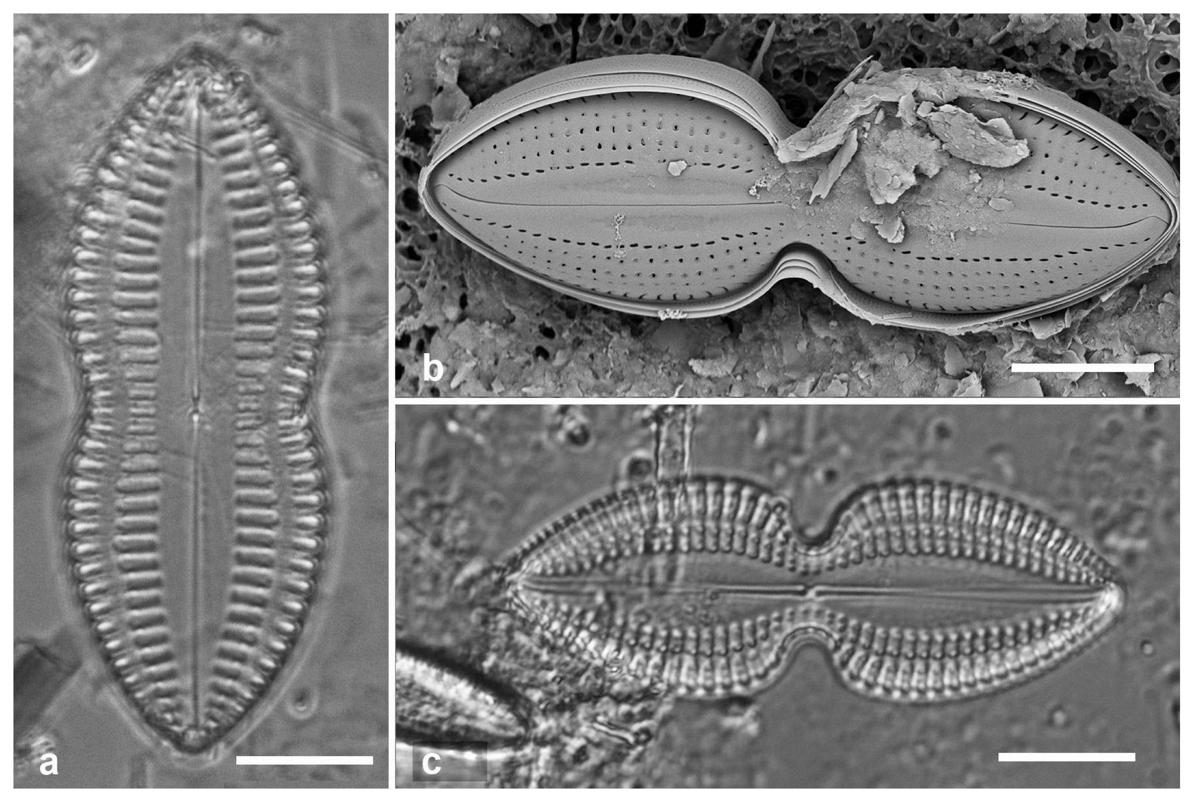
3.117. Caloneis cf. petitiana (Grunow in Cleve) Cleve 1901 [123]—Figure 41c,d
Yap samples: Y26C, Y25H-2
Dimensions: Length 88 µm, width 17 µm, striae 12 in 10 µm
3.118. Diploneis carolinensis Lobban & Witkowski 2024 [165]
Yap sample: Y34A
Dimensions: Length 19–23 μm long, greatest width 9–11 μm, striae 12 in 10 μm
(a) Caloneis egena, LM. (b) Caloneis ophiocephala, LM. (c,d) Caloneis cf. petitiana, LM at two focal planes, SEM at 15 kV showing finely porous membranes over alveoli. Scale bars: (a–c) = 10 µm, (d) = 5 µm.
Figure 41.
(a) Caloneis egena, LM. (b) Caloneis ophiocephala, LM. (c,d) Caloneis cf. petitiana, LM at two focal planes, SEM at 15 kV showing finely porous membranes over alveoli. Scale bars: (a–c) = 10 µm, (d) = 5 µm.
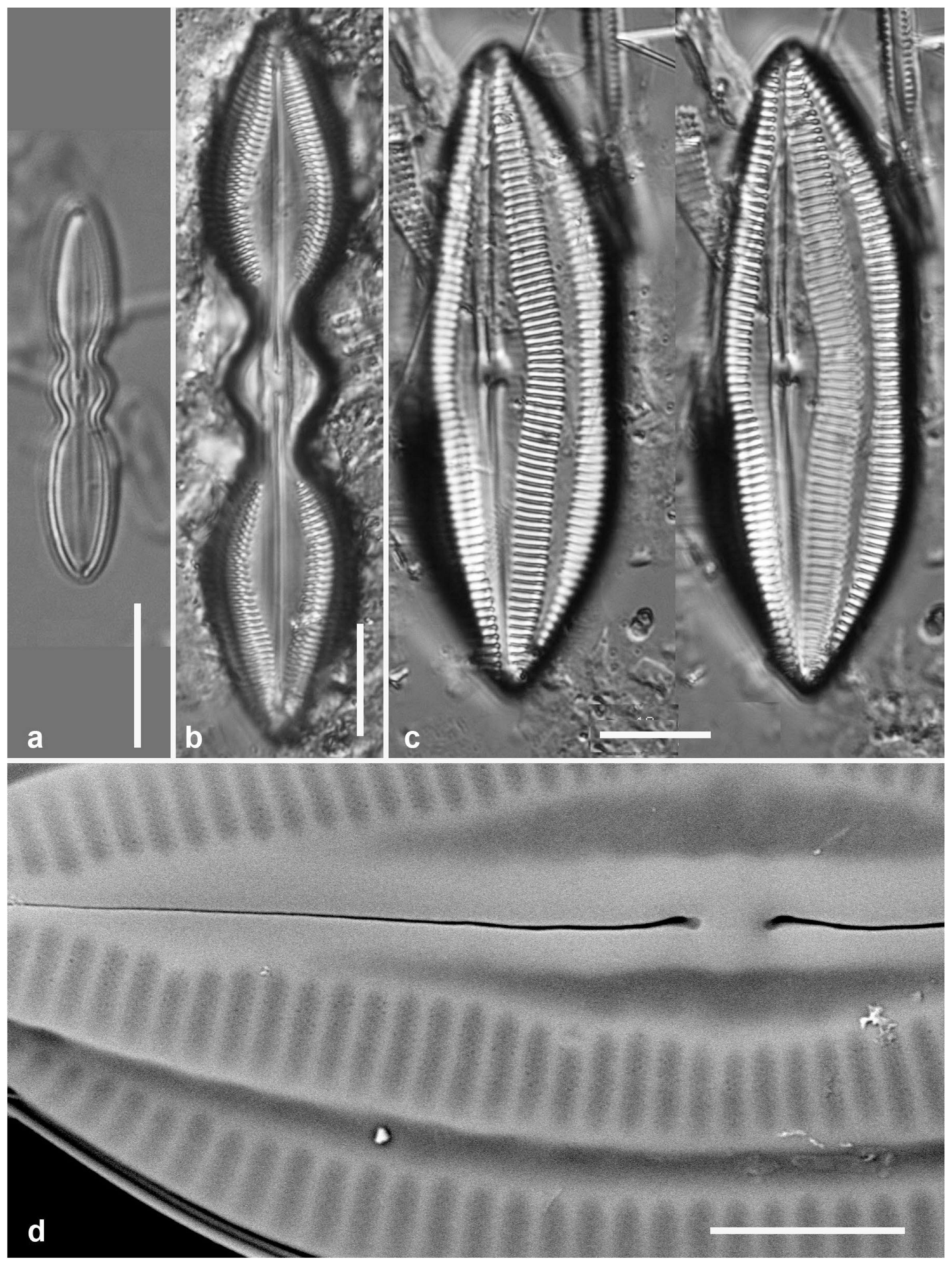
3.119. Diploneis cerebrum Pennesi, Caputo & Lobban 2017 in [166]—Figure 42
Yap samples: Y41-7, Y41-8
Dimensions: Length 55 µm, width 10 µm, striae 8 in 10 µm
3.120. Diploneis chersonensis (Grunow) Cleve 1892 in [163]—Figure 43a
Yap samples: Y36-3
Dimensions: Length 39 µm, width 16 µm, striae 12 in 10 µm
Diagnostics: Striae divided by multiple parallel longitudinal ribs, not always visible in SEM, areolae over longitudinal canals small, circular, with volae attached at single point, leaving a c-shaped slit; central canal much different from D. cerebrum: eight areolae transversely elongated and opening by convoluted slits.
Comment: This species and D. claustra (see next), in contrast to D. cerebrum, have a distinct strip over the parallel longitudinal canals, resembling a shirt placket.
3.121. Diploneis claustra Lobban & Pennesi 2017 in [166]—Figure 43b
Yap samples: Y26C
Dimensions: Length 30 µm, width 17 µm, striae 9 in 10 µm
Diagnosis: Areolae, closed by very finely porous cribra, clearly seen between virgae and vimenes, the virgae raised and angled, with support struts on the vimines (longitudinal ribs), reminiscent of a window shade, differing from D. weissflogii (A.Schmidt) Cleve and D. weissflogiopsis Lobban & Pennesi (see below) in the angled virgae and the placket-like appearance of the wall over the canals. Internal unknown but probably like D. weissflogii and D. weissflogiopsis in having one large, apparently open foramen per areola.
Diploneis cerebrum. (a) LM. (b) External valve surface, SEM. (c) Interior valve surface, again showing the longitudinal rib. (d) Detail of central part of valve with detail of “brain-like” cribra over longitudinal canal areolae; double-headed arrow shows position of longitudinal rib, arrowhead the small wave in the raphe near the central endings and single arrow the central areolae over the canal (Chuuk specimen, TK28). Scale bars: (a–c) 10 µm, (d) = 5 µm.
Figure 42.
Diploneis cerebrum. (a) LM. (b) External valve surface, SEM. (c) Interior valve surface, again showing the longitudinal rib. (d) Detail of central part of valve with detail of “brain-like” cribra over longitudinal canal areolae; double-headed arrow shows position of longitudinal rib, arrowhead the small wave in the raphe near the central endings and single arrow the central areolae over the canal (Chuuk specimen, TK28). Scale bars: (a–c) 10 µm, (d) = 5 µm.
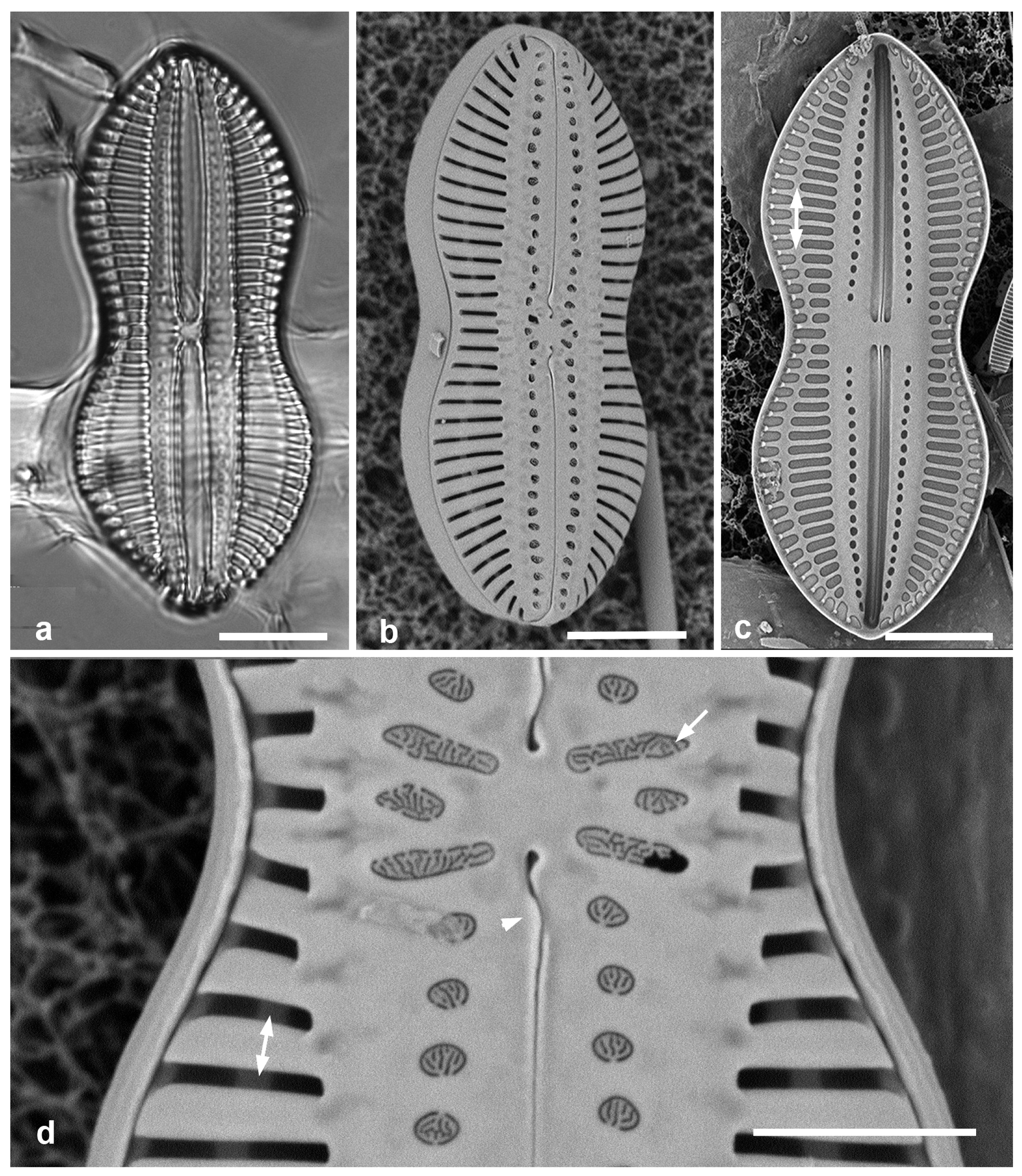
3.122. Diploneis crabro (var. crabro) Ehrenberg 1854 [167]—Figure 43c,d
Yap samples: Y18C
Dimensions: Length 84 µm, width 41 µm, striae 4.5 in 10 µm
Diploneis spp. (a) D. chersonensis, half of valve in SEM, showing flaps on raphe near central area (arrow). (b) D. claustra. (c,d) D. crabro, nominate variety. (c) LM at two focal planes showing small lunula area (arrow) and large internal foramina (arrowhead). (d) Internal SEM showing large foramina. (e) D. crabro var. excavata, LM, showing wide, excavated lunula area (arrow). Scale bars: (c–e) = 10 µm, (a,b) = 5 µm.
Figure 43.
Diploneis spp. (a) D. chersonensis, half of valve in SEM, showing flaps on raphe near central area (arrow). (b) D. claustra. (c,d) D. crabro, nominate variety. (c) LM at two focal planes showing small lunula area (arrow) and large internal foramina (arrowhead). (d) Internal SEM showing large foramina. (e) D. crabro var. excavata, LM, showing wide, excavated lunula area (arrow). Scale bars: (c–e) = 10 µm, (a,b) = 5 µm.
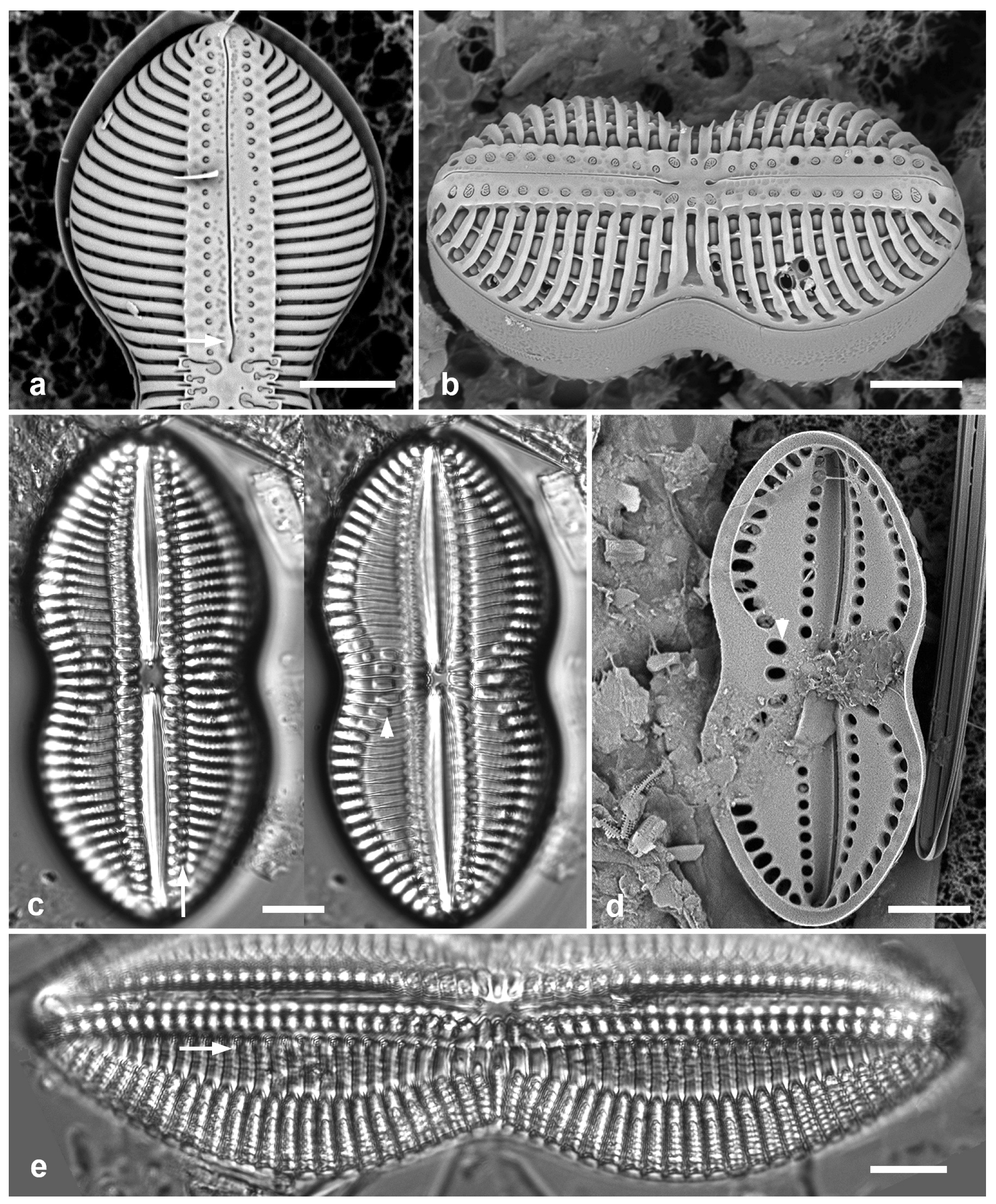
3.123. Diploneis crabro var. excavata Hustedt 1937 in [124]—Figure 43e
Yap samples: Y26C
Dimensions: Length 75–123 µm, width 25–42 µm, striae 4–6 in 10 µm
Diagnostics: Distinguished by the long excavations in the lunula area.
3.124. Diploneis craticula Pennesi, Caputo & Lobban 2017 in [166]—Figure 45a
Yap samples: Y41-7
Dimensions: Length 38 µm, width 11 µm, striae 15 in 10 µm
Diagnostics: Elliptical to slightly panduriform, transverse and longitudinal multiseriate striae; valve has only the basal silica layer, so internal aspect the same as external.
3.125. Diploneis denticulata Lobban & Prelosky, sp. nov.—Figure 44
Diagnosis: Valves lanceolate-panduriform, striae biseriate, lunula with pitted but nonperforated surface, longitudinal canals and raphe raised on flat keel with costate edge; denticulate rim between valve face and vertical mantle.
Type locality: Yap State, Yap Island, Weloy Municipality, Maa’ Mangrove, approx: 9°32′25.62″ N, 138°5′14.45″ E, adjacent to Nimpal Marine Protected Area, sample Y34A, subtidal mud from Sonneratia alba (white mangrove) pneumorrhiza, ca. 20 cm from bottom, and likely in the salt wedge. Coll. C.S. Lobban, M. Schefter & T. Gorong, 28 May 2014.
Etymology: With reference to the toothed rim between valve face and mantle.
Additional records: YAP: Y26C; PALAU: PW(2009)46; GUAM: GU55B-4.
Diploneis denticulata sp. nov., SEM except (a). Yap specimens from Y34A and (f) Y26C. (a) Holotype specimen at two focal planes in LM. (b) Exterior valve. (c) Half of same valve with natural tilt to show surface relief. (d) Oblique view of frustule, apparently in division, showing toothed rim of epitheca and inner face of daughter hypotheca. (e) Detail of inner valve surface with central raphe endings and intact hymenes covering striae. (f) Girdle view of intact frustule showing flat raphe keel, vertical mantle and hyaline cingulum. (g) Fragment of broken valve including central nodule, showing chambering in the longitudinal canal (arrow). (h) Half of valve of rounded form. Scale bars: (a) = 10 µm, (b–d,f,h) = 5 µm, (e,g) = 2 µm.
Figure 44.
Diploneis denticulata sp. nov., SEM except (a). Yap specimens from Y34A and (f) Y26C. (a) Holotype specimen at two focal planes in LM. (b) Exterior valve. (c) Half of same valve with natural tilt to show surface relief. (d) Oblique view of frustule, apparently in division, showing toothed rim of epitheca and inner face of daughter hypotheca. (e) Detail of inner valve surface with central raphe endings and intact hymenes covering striae. (f) Girdle view of intact frustule showing flat raphe keel, vertical mantle and hyaline cingulum. (g) Fragment of broken valve including central nodule, showing chambering in the longitudinal canal (arrow). (h) Half of valve of rounded form. Scale bars: (a) = 10 µm, (b–d,f,h) = 5 µm, (e,g) = 2 µm.
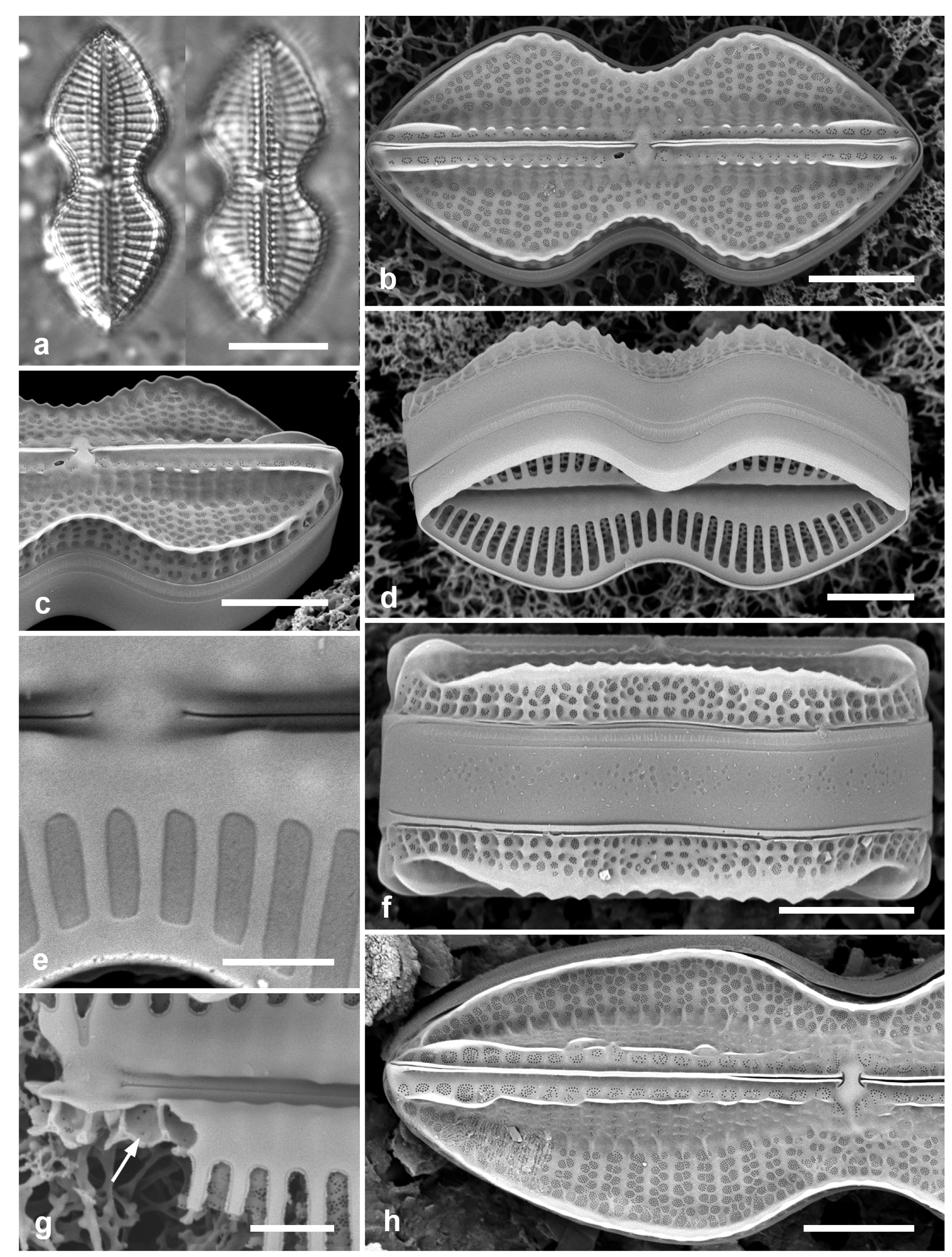
Diploneis spp. (a) D. craticula, SEM exterior. (b–d) D. papula, external views of frustules, oblique view showing domed cribra of areolae between virgae (arrow). (e) D. nitescens, SEM of valve exterior. (f) D. smithii, SEM of valve exterior. (g) D. smithii var. rhombica, LM. (h) D. suborbicularis, LM. Scale bars: (e–h) = 10 µm, (a–d) = 5 µm.
Figure 45.
Diploneis spp. (a) D. craticula, SEM exterior. (b–d) D. papula, external views of frustules, oblique view showing domed cribra of areolae between virgae (arrow). (e) D. nitescens, SEM of valve exterior. (f) D. smithii, SEM of valve exterior. (g) D. smithii var. rhombica, LM. (h) D. suborbicularis, LM. Scale bars: (e–h) = 10 µm, (a–d) = 5 µm.
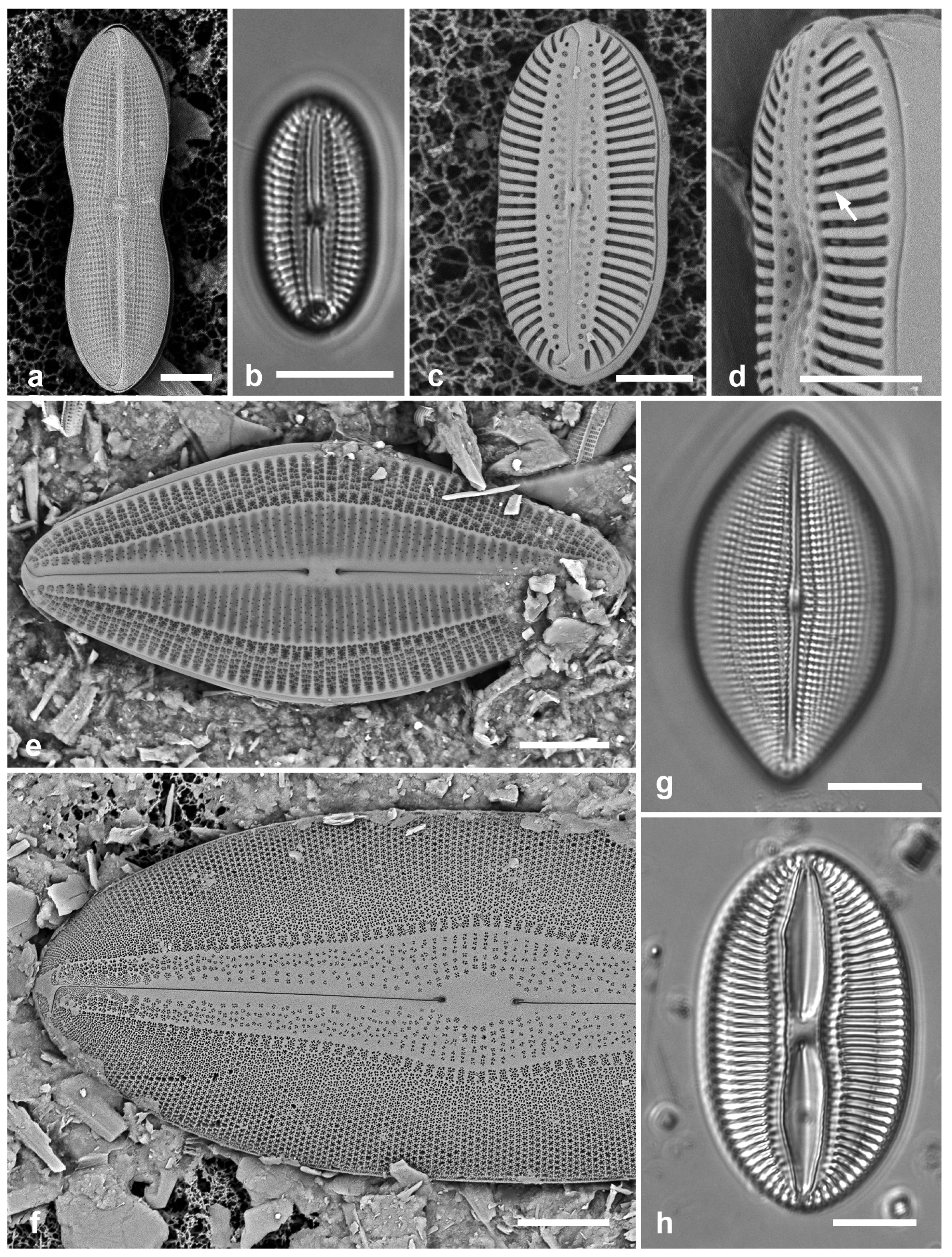
3.126. Diploneis nitescens (W.Gregory) Cleve 1894 [162]—Figure 45e
Yap samples: Y34H, Y34F, Y37-8, Y39A, Y26A, Y26B, Y26C
Dimensions: Length 43–66 µm, width 19–28 µm, striae 8–9 in 10 µm
Diagnostics: Distinguished from D. smithii (Brébisson) Cleve (see below) by the greater proportion of valve face over the longitudinal canals, i.e., 1/3 vs. 1/4–1/3.
3.127. Diploneis papula (A.Schmidt) Cleve 1894 [162]—Figure 45b–d
Yap samples: Y37-7, Y36-1, Y41-7, Y41-8
Dimensions: Length 21–24 µm, width 11–12 µm, striae 12–13 in 10 µm
3.128. Diploneis smithii (Brébisson) Cleve 1894 [162]—Figure 45f
Yap samples: Y25H-1, Y41-7, -8
Dimensions: Length 35–99 µm, width 15–41 µm, striae 8–10 in 10 mm
3.129. Diploneis smithii var. rhombica Mereschkowsky 1902 [171]—Figure 45g
Yap samples: Y33A
Dimensions: Length 45 µm, width 24 µm, striae 11 in 10 mm
3.130. Diploneis suborbicularis (W.Gregory) Cleve 1894 [162]—Figure 45h
Yap samples: Y25H-1, Y25H-2
Dimensions: Length 45 µm, width 26 µm, striae 8 in 10 mm
3.131. Diploneis weissflogii (A.Schmidt) Cleve 1894 [162]—Figure 46a,b
Yap samples: Y25H-1, Y25H-2, Y26B, Y37-8
Dimensions: Length 26–30 µm, width 11 µm, striae 9–10 in 10 µm
3.132. Diploneis weissflogiopsis Lobban & Pennesi 2017 in [166]—Figure 46c,d
Yap samples: Y18C
Dimensions: Length 27–43 µm, width 9–14 µm, striae 10–12 in 10 µm
(a–d) Diploneis weissflogii vs. D. weissflogiopsis, SEM. (a,b) D. weissflogii. (a) Internal view showing single foramen on each side of the central area (in rectangle), foramina at apex of canals (arrows), and hyaline copulae. (b) External valve face showing single modified stria between central raphe endings (in rectangle). (c,d) D. weissflogiopsis. (c) Valve showing three modified striae between raphe endings (in rectangle); the higher striae density is also evident in the comparison. (d) Detail of central area in slightly oblique view with the central dimple more evident (arrow). (e) Navicula consors. SEM. Scale bars: (e) =10 µm, (a–d) = 5 µm.
Figure 46.
(a–d) Diploneis weissflogii vs. D. weissflogiopsis, SEM. (a,b) D. weissflogii. (a) Internal view showing single foramen on each side of the central area (in rectangle), foramina at apex of canals (arrows), and hyaline copulae. (b) External valve face showing single modified stria between central raphe endings (in rectangle). (c,d) D. weissflogiopsis. (c) Valve showing three modified striae between raphe endings (in rectangle); the higher striae density is also evident in the comparison. (d) Detail of central area in slightly oblique view with the central dimple more evident (arrow). (e) Navicula consors. SEM. Scale bars: (e) =10 µm, (a–d) = 5 µm.
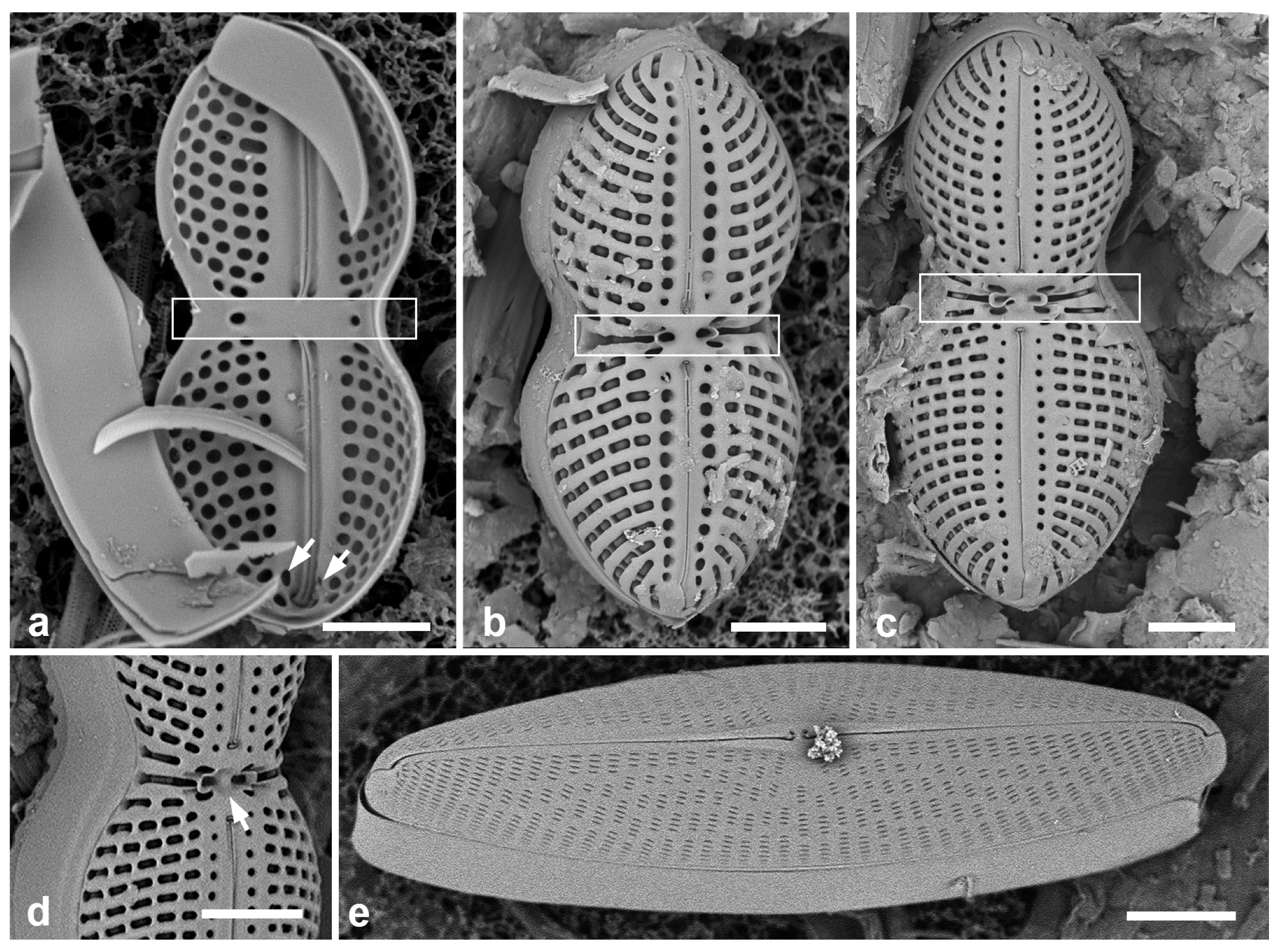
3.133. Cymatoneis sulcata (Greville) Cleve 1894 [162]—Figure 47a,b
Additional Yap samples: Y25B
Dimensions: Length 32–50 µm, width 16 µm, striae 10–11 in 10 µm
Cymatoneis spp., SEM. (a,b) C. sulcata, Palau specimens (PW46). Valve and girdle views showing strongly developed longitudinal ribs and hyaline cingulum. (c) C. belauensis, sp. nov. from Palau (PW46) valve view, showing longitudinal ridges with hyaline outer sides and ribs enclosing much of raphe. (d). Same valve naturally tilted, showing elevations of the valve. Scale bars: (a–c) = 10 µm, (d) = 5 µm.
Figure 47.
Cymatoneis spp., SEM. (a,b) C. sulcata, Palau specimens (PW46). Valve and girdle views showing strongly developed longitudinal ribs and hyaline cingulum. (c) C. belauensis, sp. nov. from Palau (PW46) valve view, showing longitudinal ridges with hyaline outer sides and ribs enclosing much of raphe. (d). Same valve naturally tilted, showing elevations of the valve. Scale bars: (a–c) = 10 µm, (d) = 5 µm.
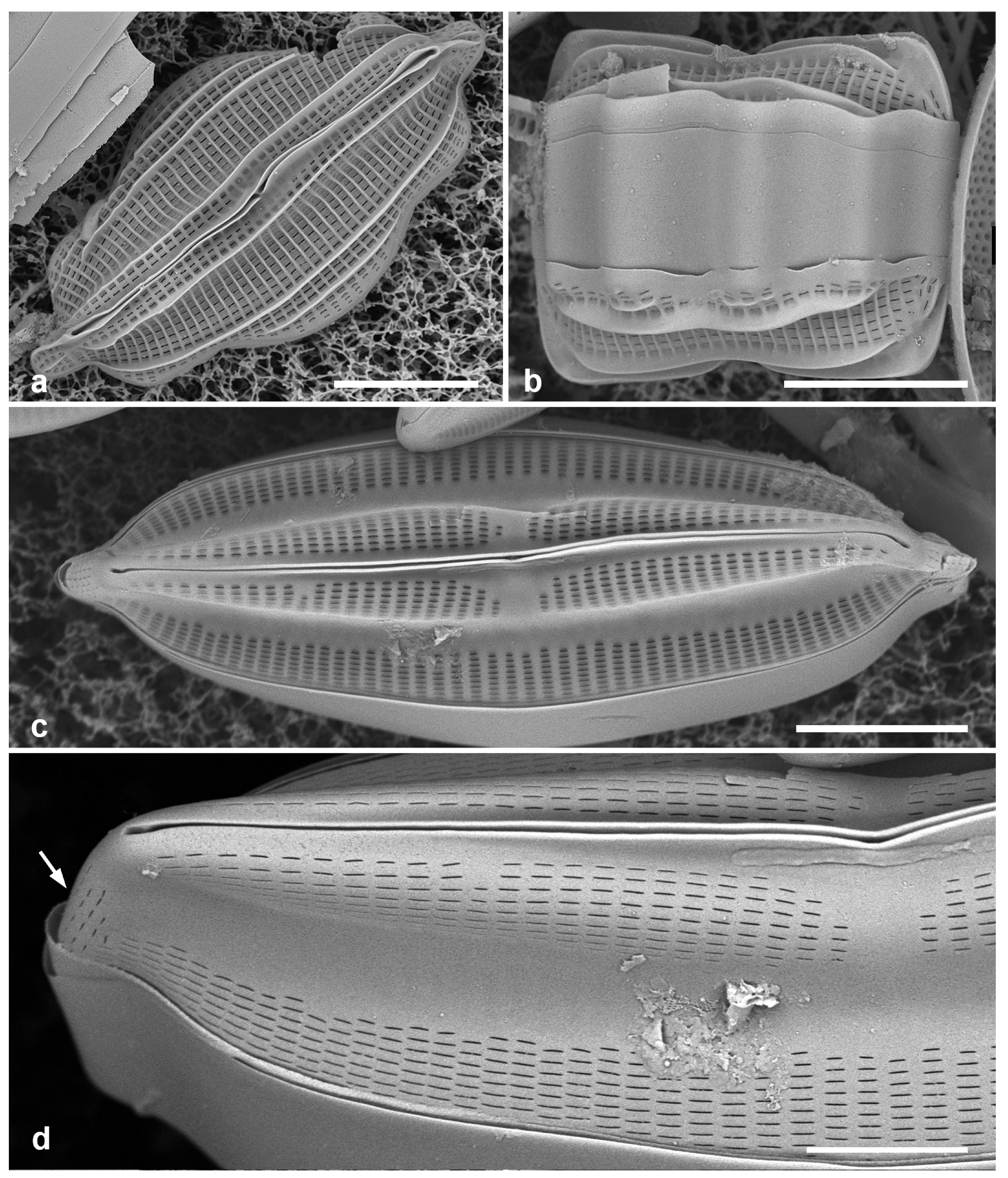
3.134. Cymatoneis belauensis Lobban, sp. nov.—Figure 47c,d and Figure 48a–c
Diagnosis: Valve face with lateral sterna joined by central area, vimenes not thickened into transverse costae, striae densely areolate, raphe branches partially bordered by ribs.
Type locality: PALAU: Babeldaob Island, Ngaremlengui State, dock at Bkulangriil, 07°31.488′ N, 134°29.966′ E, sand sample rich in Carinasigma, sample PW2009-46. Coll. 11 April 2009, C.S. Lobban and M. Schefter.
Etymology: Named after the country where it was found.
3.135. Cymatoneis yapensis Lobban, sp. nov.—Figure 48d
Diagnosis: Differing from C. belauensis in smaller size and sparser areolae in the striae.
Type locality: Yap State, Yap Island, Weloy Municipality, Maa’ Mangrove, approx: 9°32′25.62″ N, 138°5′14.45″ E, adjacent to Nimpal Marine Protected Area, sample Y34A, subtidal mud from Sonneratia alba (white mangrove) pneumorrhiza, ca 20 cm from bottom, and likely in the salt wedge. Coll. C.S. Lobban, M. Schefter & T. Gorong, 28 May 2014
Etymology: Named after the state where it was found.
Cymatoneis spp., cont. (a–c) C. belauensis, internal aspects of a valve. (a) Valve as found, showing valve depth from outside and outer zone of the striae. (b,c) Same valve tilted 45°; (b) showing whole valve, (c) detail of central area with small central nodule. (d) C. yapensis, sp. nov. from Yap (Y34A). Scale bars: (a,b,d) = 10 µm, (c) = 5 µm.
Figure 48.
Cymatoneis spp., cont. (a–c) C. belauensis, internal aspects of a valve. (a) Valve as found, showing valve depth from outside and outer zone of the striae. (b,c) Same valve tilted 45°; (b) showing whole valve, (c) detail of central area with small central nodule. (d) C. yapensis, sp. nov. from Yap (Y34A). Scale bars: (a,b,d) = 10 µm, (c) = 5 µm.

3.136. Navicula consors A.W.F.Schmidt 1876 in [162]—Figure 45e
Yap samples: Y37-8, Y37-7, Y37-8, Y41-7
Dimensions: Length 64–73 µm, width 16–18 µm, striae 7 in 10 µm
3.137. Navicula plicatula Grunow 1894 in [162]—Figure 49a,b
Yap samples: Y26C
Dimensions: Length 95 µm, width 32 µm, striae 15 in 10 µm
3.138. Navicula tsukamotoi (Sterrenburg & Hinz) Yuhang Li & Kuidong Xu 2017 in [173]—Figure 49c
Yap samples: Y25H-1, Y25H-2, Y26C, Y37-7, Y37-8, Y36-1, Y41-7
(a,b) Navicula plicatula SEM external and internal aspects of valve. (c) Navicula tsukamotoi valve external, SEM. Scale bars = 10 µm.
Figure 49.
(a,b) Navicula plicatula SEM external and internal aspects of valve. (c) Navicula tsukamotoi valve external, SEM. Scale bars = 10 µm.
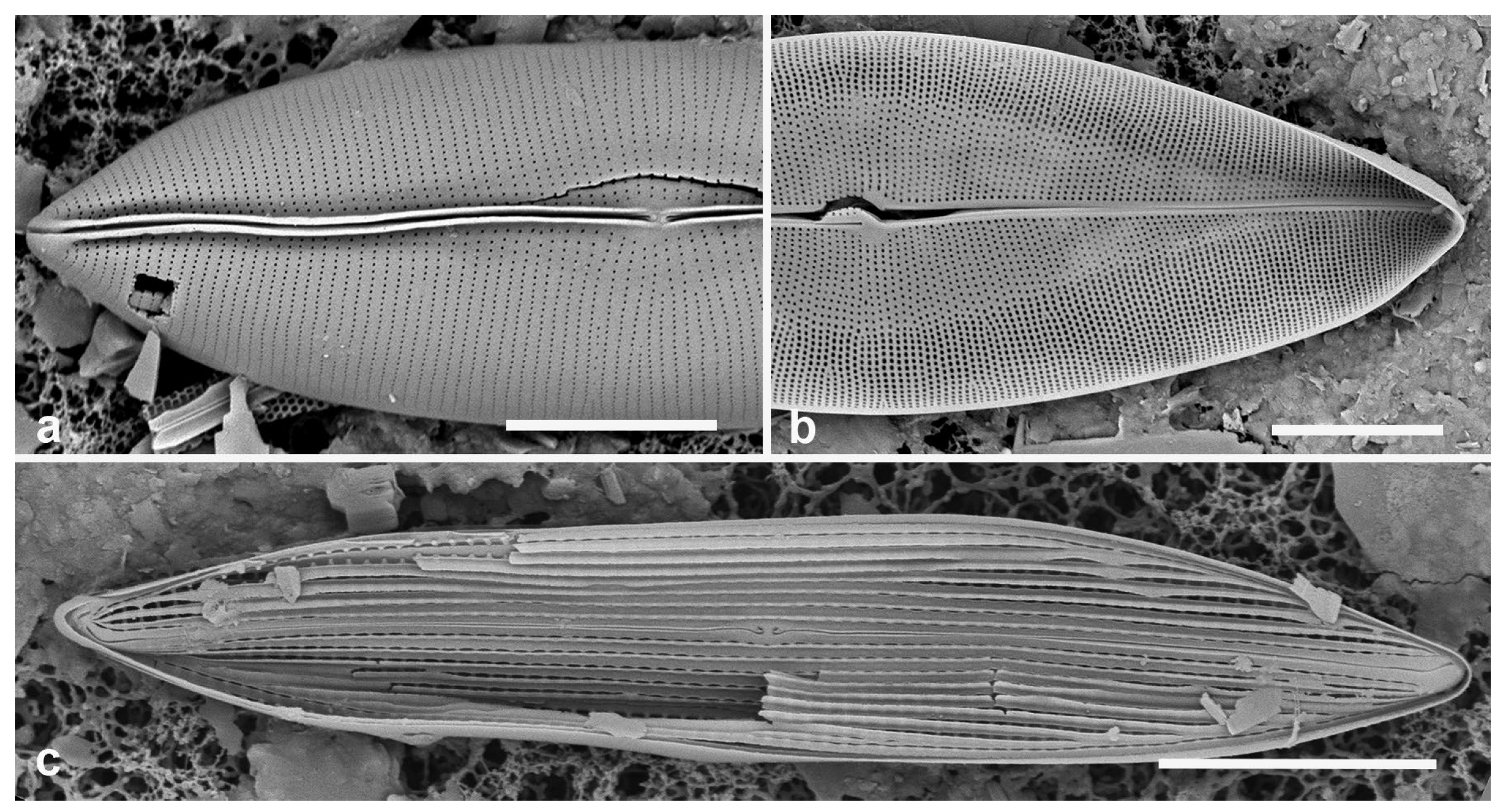
Dimensions: Length 59–65 µm, width 11 µm, striae 16–17 in 10 µm
3.139. Trachyneis aspera (Ehrenberg) Cleve 1894 [162]—Figure 50a
Yap samples: Y25H-2, Y26C, Y37-7, Y37-8, Y36-1
Dimensions: Length 45–70 µm, width 13–16 µm, striae 13–16 in 10 µm
3.140. Trachyneis velata (A. Schmidt) Cleve 1894 [162]—Figure 50b
Yap samples: Y25H-1, Y37-7
Plagiotropidaceae D.G. Mann
3.141. Plagiotropis lepidoptera (W.Gregory) Kuntze 1898 [176]—Figure 50c–g and Figure 51
Yap samples: Y25H-1, Y41-7, -8
Dimensions: Length 59 µm, width 14 µm, striae 22–23 in 10 µm
(a) Trachyneis aspera. (b) Trachyneis velata. (c–g) Plagiotropis lepidoptera. (c) LM at two focal planes. (d,e) SEM of external valve faces showing major and minor sides and paired areolae (arrow, Figure 50e). (f,g) Central area with enlargement of central nodule. Scale bars: (a–d) = 10 µm, (f) = 5 µm, (e) = 2 µm, (g) = 1 µm.
(a) Trachyneis aspera. (b) Trachyneis velata. (c–g) Plagiotropis lepidoptera. (c) LM at two focal planes. (d,e) SEM of external valve faces showing major and minor sides and paired areolae (arrow, Figure 50e). (f,g) Central area with enlargement of central nodule. Scale bars: (a–d) = 10 µm, (f) = 5 µm, (e) = 2 µm, (g) = 1 µm.
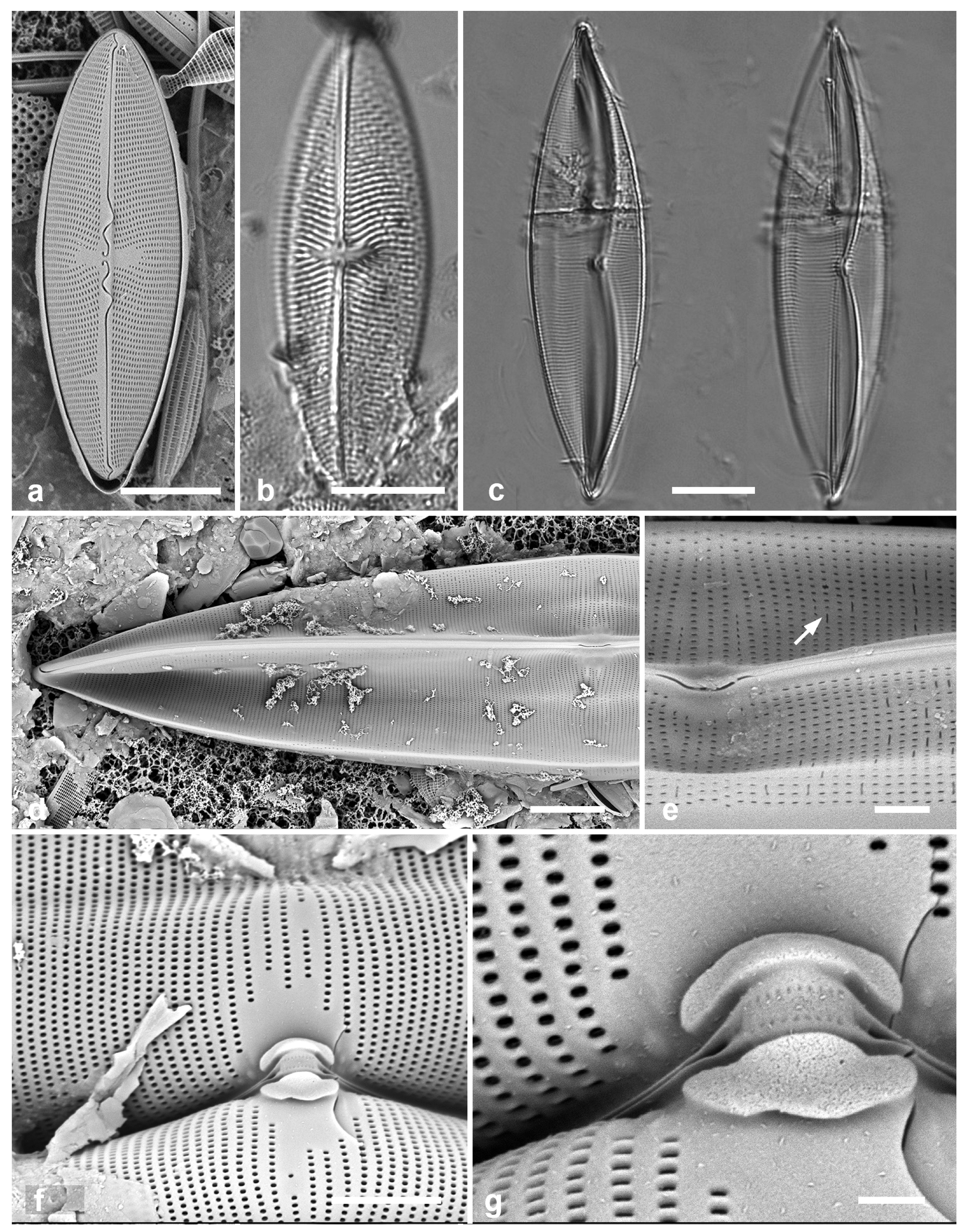
Morphological characters in Plagiotropis across Micronesia. (a) Unidentified species with scuta (arrows) (Y34H). (b) Portion of a valve showing double wall with external silica flap (arrow) forming the scutum (PW1990-47). (c) Portion of a valve showing single wall pleated to produce the scutum (arrow) (GU68D-1B). (d) Specimen with conopeum over central raphe endings (arrow) (Y45-2). Scale bars: (a,d) = 10 µm, (b,c) = 2 µm.
Figure 51.
Morphological characters in Plagiotropis across Micronesia. (a) Unidentified species with scuta (arrows) (Y34H). (b) Portion of a valve showing double wall with external silica flap (arrow) forming the scutum (PW1990-47). (c) Portion of a valve showing single wall pleated to produce the scutum (arrow) (GU68D-1B). (d) Specimen with conopeum over central raphe endings (arrow) (Y45-2). Scale bars: (a,d) = 10 µm, (b,c) = 2 µm.
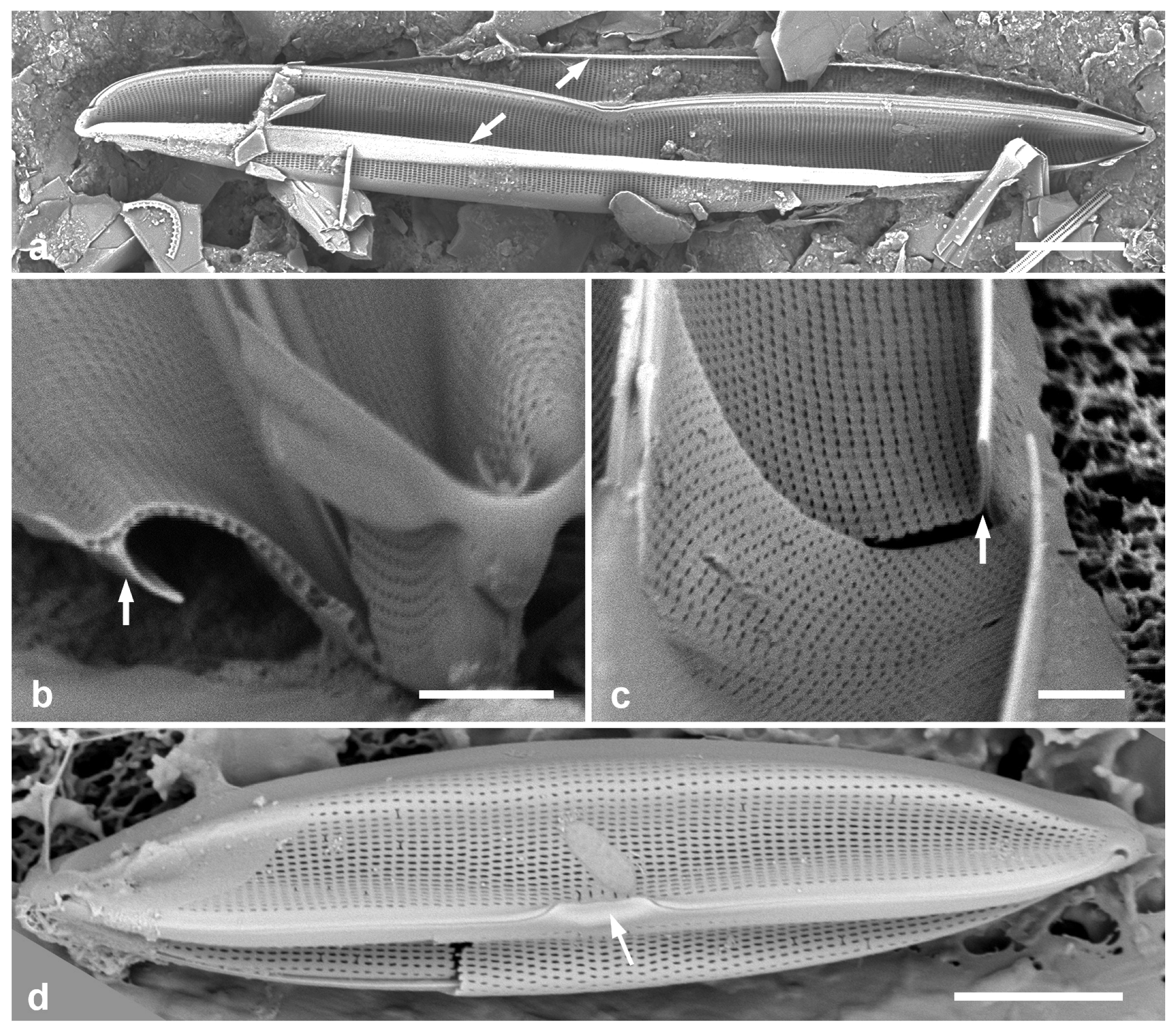
Pleurosigmataceae Mereschkowsky
3.142. Pleurosigma simulacrum Lobban & F.A.S. Sterrenberg in [21]—Figure 52a,b
Additional Yap samples: Y26B, Y26C, Y42-1
Dimensions: Length 110–133 μm, width 13–14 μm, transverse striae 26 in 10 μm
3.143. Rhoicosigma parvum Hein & Lobban 2015 [177]—Figure 52c,d
Yap samples: Y37-8, Y36-1, Y41-7
Dimensions: Length 48 μm, width 8 μm, transverse striae 27 in 10 μm
Diagnostics: In this genus, valves bent across the pervalvar axis, the concave valve with sigmoid raphe, convex with nearly straight raphe. This species is smaller than R. compactum (Greville) Grunow reported for Micronesia.
(a,b) Pleurosigma simulacrum, Yap voucher specimen in SEM at two magnifications. (c,d) Rhoicosigma parvum. Convex valve with straight raphe in LM, concave valve with sigmoid raphe in SEM. (e–g) Schizostauron cf. trachyderma. (e) Raphe valve in LM. (f) Sternum valve in LM. (g) Sternum valve internal aspect in SEM. Scale bars: (a) = 20 µm, (c–g) = 10 µm, (b) = 5 µm.
Figure 52.
(a,b) Pleurosigma simulacrum, Yap voucher specimen in SEM at two magnifications. (c,d) Rhoicosigma parvum. Convex valve with straight raphe in LM, concave valve with sigmoid raphe in SEM. (e–g) Schizostauron cf. trachyderma. (e) Raphe valve in LM. (f) Sternum valve in LM. (g) Sternum valve internal aspect in SEM. Scale bars: (a) = 20 µm, (c–g) = 10 µm, (b) = 5 µm.

Stauroneidaceae D.G. Mann
3.144. Schizostauron cf. trachyderma (F.Meister) Górecka & Riaux-Gobin 2021 in [178]—Figure 52e–g
Yap samples: Y25H-2
Dimensions: Length 32 µm, width 16 µm, striae 10 in 10 µm
THALASSIOPHYSALES D.G. Mann
Catenulaceae Mereschkowsky
3.145. Amphora arenaria Donkin 1858 [179]—Figure 53a
Yap samples: Yap samples: Y25H-1
Dimensions: Length 72 µm, width 11, striae ca. 25 in 10 µm
3.146. Amphora bigibba Grunow 1875 in [162]—Figure 53b,c
Amphora spp. (a) A. arenaria, LM. (b,c) A. bigibba SEM of frustules, arrows showing rows of apically elongate areolae near raphe ledge. (d,e) A. bigibba var. interrupta. SEM of frustule in ventral view and isolated valve, external aspect. Scale bars: (a) = 10 µm, (b–e) = 5 µm.
Figure 53.
Amphora spp. (a) A. arenaria, LM. (b,c) A. bigibba SEM of frustules, arrows showing rows of apically elongate areolae near raphe ledge. (d,e) A. bigibba var. interrupta. SEM of frustule in ventral view and isolated valve, external aspect. Scale bars: (a) = 10 µm, (b–e) = 5 µm.
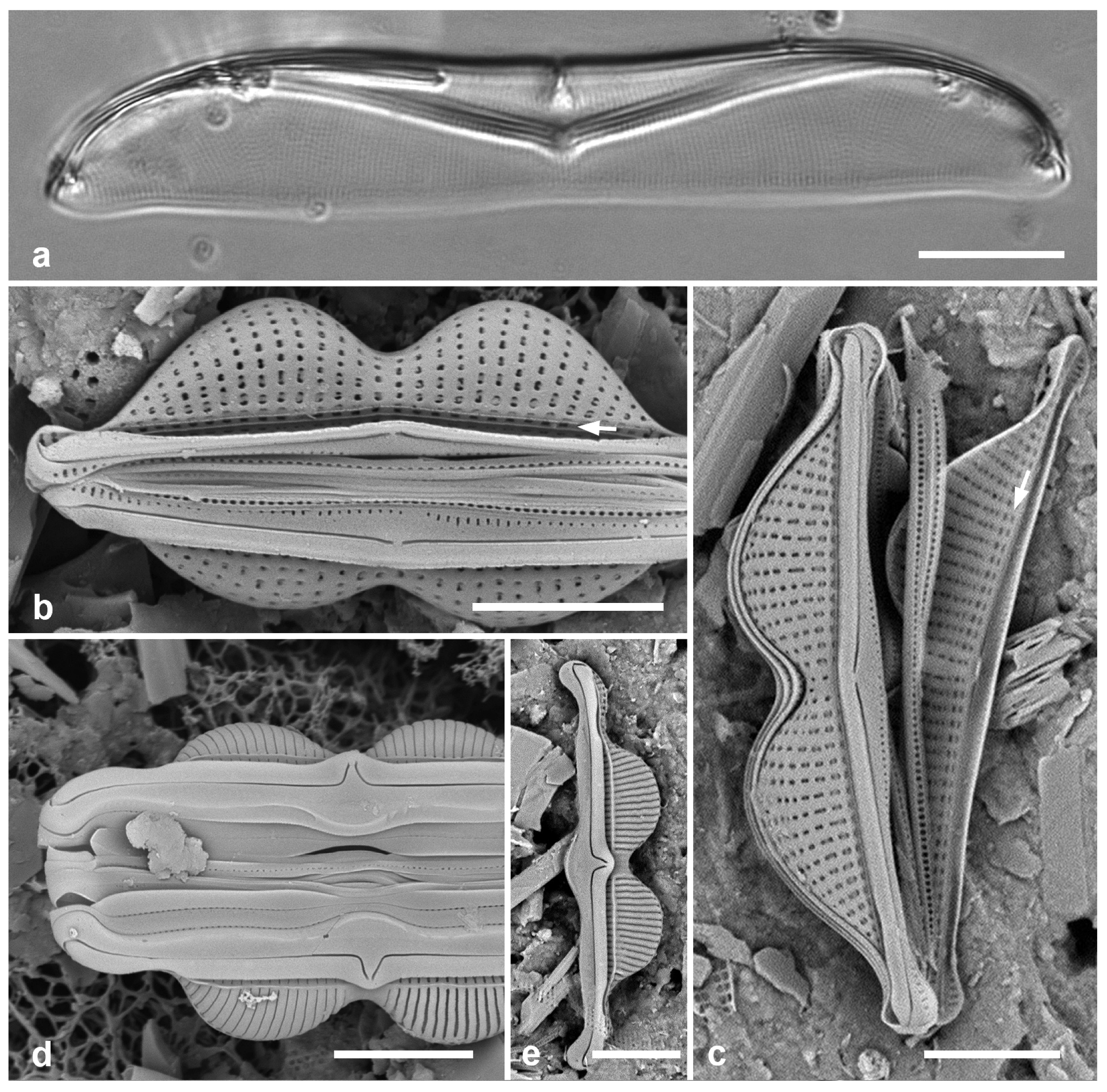
Yap samples: Y25H-1, Y26B, Y37-8, Y41-7
Dimensions: Length 12–20 µm, width 4–5, dorsal striae 19–20 in 10 µm, ventral 31–36 in 10 µm
Diagnostics: Small cells with rostrate apices and a strong constriction in the middle of the dorsal side, hence the name bigibba.
3.147. Amphora bigibba var. interrupta (Grunow) Cleve 1895 [183]—Figure 53d,e
Yap samples: Y26C
Dimensions: Length 23 µm, width 4 µm, dorsal striae 27 in 10 µm
Diagnostics: Bigibbous valve with long protracted apices, strongly bent central raphe endings, and break in dorsal striae at center of valve. Dorsal areolae hidden by overlying virgae development leaving slit; ventral areolae absent. There is a longitudinal break in the striae near the raphe ledge.
3.148. Amphora hyalina Kützing 1844 [37]
Dimensions: Length 40–45 µm, width 8 µm, striae 28–30 in 10 µm.
3.149. Amphora immarginata Nagumo 2003 [185]—Figure 54a
Yap samples: Y37-8, Y41-7
Dimensions: Length 26–43 µm, width 8–10 µm, dorsal and ventral striae 17–18 in 10 µm
3.150. Amphora obtusa W.Gregory 1857 [186]—Figure 54b
Yap samples: Y26C
Dimensions: Length 49–106 µm, width 14–16 µm, dorsal and ventral striae 19–23 in 10 µm
(a) Amphora obtusa, LM. (b) Amphora immarginata, SEM valve external. (c) Amphora ostrearia var. vitrea, SEM valve external. (d,e) Amphora proteus, LM, vale and frustule. (f) Amphora spectabilis, LM. (g) Amphora subhyalina, SEM of frustule. Scale bars = 10 µm.
Figure 54.
(a) Amphora obtusa, LM. (b) Amphora immarginata, SEM valve external. (c) Amphora ostrearia var. vitrea, SEM valve external. (d,e) Amphora proteus, LM, vale and frustule. (f) Amphora spectabilis, LM. (g) Amphora subhyalina, SEM of frustule. Scale bars = 10 µm.
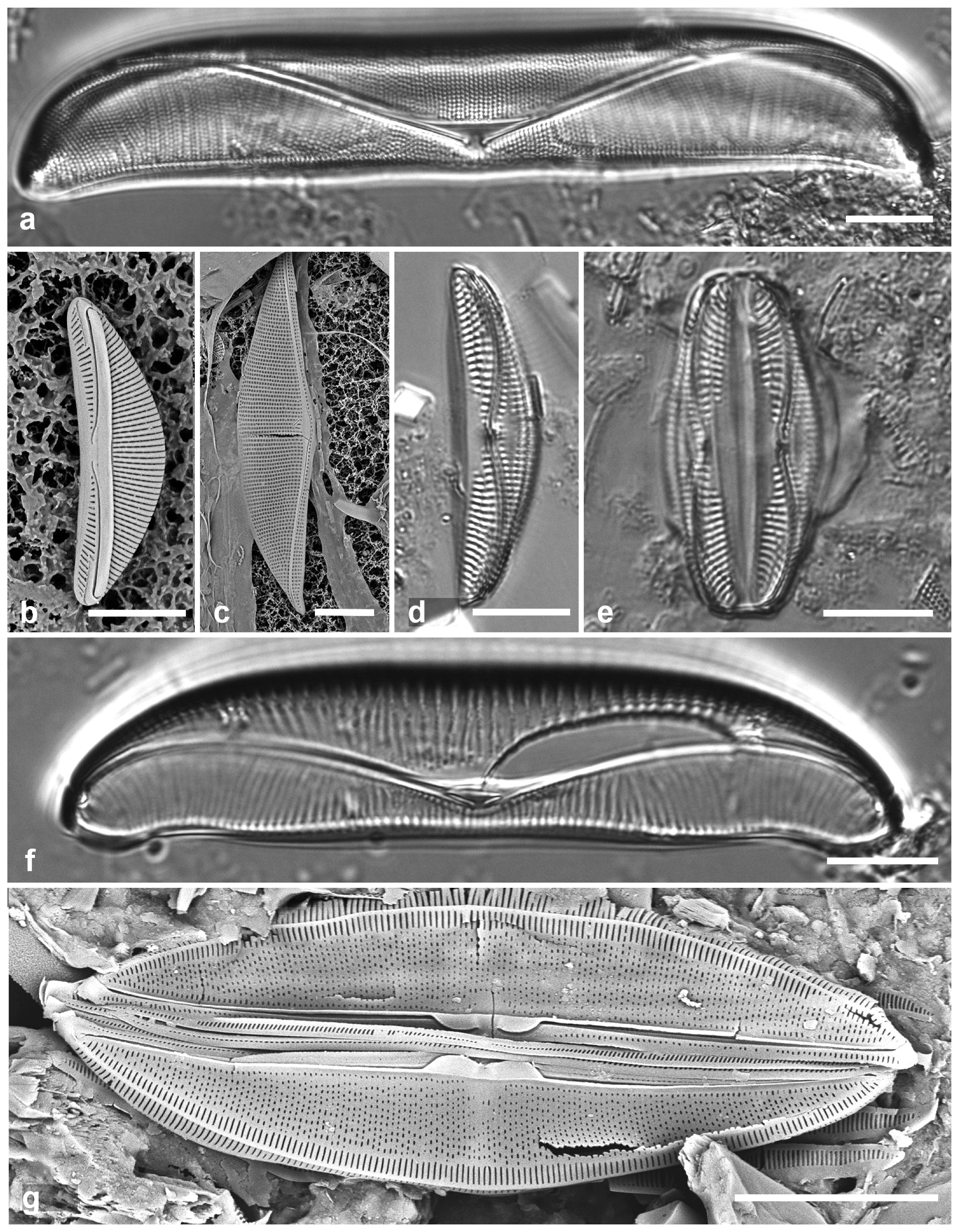
3.151. Amphora ostrearia var. vitrea (Cleve) Cleve 1895 [183]—Figure 54c
Yap samples: Y41-7
Dimensions: Length 64 µm, width 14 µm, dorsal and ventral striae 15 in 10 µm.
3.152. Amphora cf. proteus W. Gregory 1857 [151]—Figure 54d,e
Yap samples: Y25H-1, Y18B
Dimensions: Length 31–36 µm, width 8 µm, dorsal striae 15 in 10 µm, ventral striae 12 in 10 µm
3.153. Amphora spectabilis W.Gregory 1857 [151]—Figure 54f
Yap samples: Y26C
Dimensions: Length 50–78 µm, width 14–17 µm, dorsal striae 7 in 10 µm, ventral striae 14 in 10 µm
3.154. Amphora subhyalina Podzorski & Håkansson 1987 [41]—Figure 54g
Synonym: A. insulana Stepanek & Kociolek
Yap samples: Y26C, Y41-8
Dimensions: Length 27–42 µm, width 6–7 µm, dorsal striae 34–36 in 10 µm, ventral striae 37 in 10 µm
3.155. Halamphora exigua (W.Gregory) Levkov 2009 [188]
3.156. Halamphora turgida (W.Gregory) Levkov 2009 [188]
Thalassiophysidaceae D.G. Mann
3.157. Thalassiophysa hyalina (Greville) Paddock & Sims 1981 [189]—Figure 55a,b
Yap samples: Y25H-2
Dimensions: Length 112 µm, width 30 µm, striae ca. 80 in 10 µm
(a,b) Thalassiophysa hyalina. (a) Valve in LM. (b) Center of valve exterior in SEM. (c) Undatella lineata valve in LM. (d) “Bacillaria paradoxa” Group B, SEM showing characteristic T-shaped terminal raphe ending (arrow). (e,f) Cymatonitzschia marina, frustules in LM and SEM. Scale bars: (a,c,e,f) = 10 µm, (b,d) = 5 µm.
Figure 55.
(a,b) Thalassiophysa hyalina. (a) Valve in LM. (b) Center of valve exterior in SEM. (c) Undatella lineata valve in LM. (d) “Bacillaria paradoxa” Group B, SEM showing characteristic T-shaped terminal raphe ending (arrow). (e,f) Cymatonitzschia marina, frustules in LM and SEM. Scale bars: (a,c,e,f) = 10 µm, (b,d) = 5 µm.
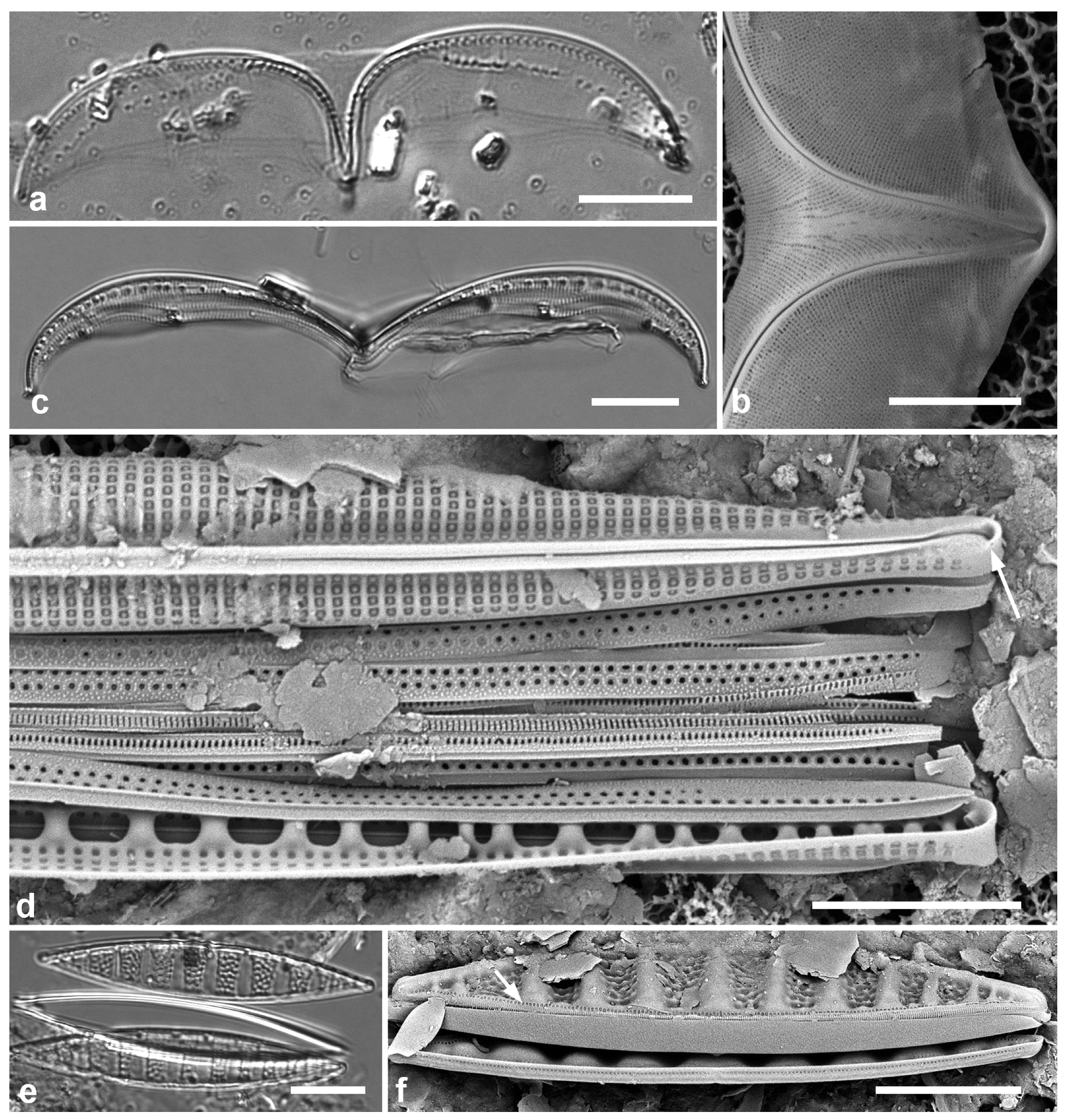
3.158. Undatella lineata (Greville) Paddock & Sims 1980 [190]—Figure 55c
Yap samples: Y26C, Y41-7
3.159. “Bacillaria paradoxa” Group B sensu Schmid 2007 [191]—Figure 55d
Yap samples: Y25H-1, Y37-8
Dimensions: Length 76–113 µm, width 4–5 µm, striae 24 in 10 µm, fibulae 7–8 in 10 µm
3.160. Cymatonitzschia marina (Lewis) Simonsen 1974 [122]—Figure 55e,f
Yap samples: Y25H-2, Y26B, Y26C
Dimensions: Length 43–67 µm, width 8–9 µm
3.161. Gomphotheca marciae Lobban & Prelosky 2022 [22]
Dimensions: Length 348–624 μm, width tapering uniformly from 9.5–12.3 μm near apex to 4.2–4.7 μm near base; striae 35–45 in 10 μm, fibulae 5.5–6.5 in 10 μm
Comments: This large and robust species is from mangrove habitat.
Homoeocladia spp.
Homoeocladia spp. Yap vouchers, SEM. (a) H. schefteropsis, showing areolae in peri-raphe zone (black arrows) and squiggly areolae on copulae (white arrow). (b) H. coacervata, showing areolae in peri-raphe zone (black arrow) and stacked slits on copulae (white arrow). (c) H. micronesica, showing no areolae in peri-raphe zone (black arrow). A row of small pores is present on the apex in all three species (arrowheads). Scale bars = 2 µm.
Figure 56.
Homoeocladia spp. Yap vouchers, SEM. (a) H. schefteropsis, showing areolae in peri-raphe zone (black arrows) and squiggly areolae on copulae (white arrow). (b) H. coacervata, showing areolae in peri-raphe zone (black arrow) and stacked slits on copulae (white arrow). (c) H. micronesica, showing no areolae in peri-raphe zone (black arrow). A row of small pores is present on the apex in all three species (arrowheads). Scale bars = 2 µm.
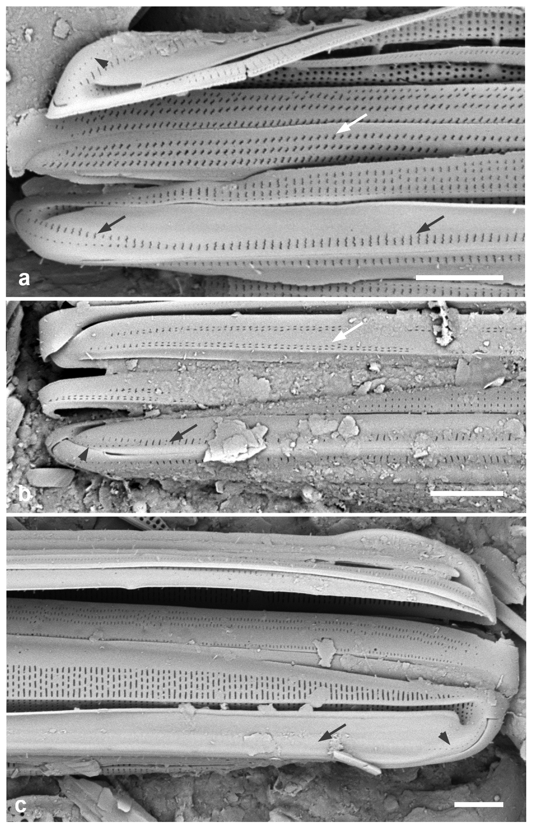
3.162. Homoeocladia celaenopsis Lobban, Sison and Ashworth 2023 [24]
Dimensions: Length 46–58 µm, width 6 µm, striae 50 in 10 µm, fibulae 3–4 in 10 µm
Diagnostics: Moderate length, lanceolate, areolae S-shaped, weakly spathulate, two rows of S-shaped pores along each side of the peri-raphe zone.
3.163. Homoeocladia radiata Lobban, Sison, and Ashworth 2023 [24]
Dimensions: Length 39–51 µm, width 6 µm, striae 50 in 10 µm
Diagnostics: Moderate length, lanceolate, striae strongly radiate towards apices.
3.164. Homoeocladia vittaelatae Lobban, Sison and Ashworth 2023 [24]
Dimensions: Length 89–98 µm, width 11 µm, striae 47 in 10 µm, fibulae 3.5 in 10 µm
Diagnostics: Long, linear, nonspathulate, copulae deep and densely perforated (visible in LM).
3.165. Homoeocladia schefteropsis Lobban, Sison and Ashworth 2023 [24]—Figure 56a
Dimensions: Length 27–40 µm, width 4.5 µm, striae 55–56 in 10 µm, fibulae 5 in 10 µm
Diagnostics: Moderate length, lanceolate, indistinguishable from H. coacervata (next taxon) except by sigmoid girdle band areolae.
3.166. Homoeocladia coacervata Lobban, Sison and Ashworth 2023 [24]—Figure 56b
Dimensions: Length 30–39 µm, width 3–4 µm, striae 55 in 10 µm, fibulae 6–7 in 10 µm
Diagnostics: Moderate length, lanceolate, areolae on the copulae in short stacks of straight slits.
3.167. Homoeocladia micronesica Lobban, Sison and Ashworth 2023 [24]—Figure 56c
Dimensions: Length 86 µm, width 7 µm, striae 44–46 in 10 µm, fibulae 4 in 10 µm
Diagnostics: Long, linear, spathulate, differing from others of similar size and shape in areolae character and position
3.168. Homoeocladia martiana C. Agardh 1827 [194]—Figure 57a,b
Yap samples: Y26C
Dimensions: Length about 230 µm, entire specimens not observed in this sediment sample; width 3.5 µm, striae 30 in 10 µm, fibulae 6–7 in 10 µm
Comments: This tube-dwelling species that can form stout colonies up to 10 cm length.
3.169. Homoeocladia tarangensis (Lobban) Lobban & Ashworth 2022 [197]
Dimensions: Length 109–194 µm, width 10–12 µm, striae 46 in 10 µm, fibulae irregular, 2 in 10 µm.
Diagnostics: Valves long, spathulate in girdle view because of apical keel extensions; valve linear with rib along the edge of the valve depression, small pores in peri-raphe zone, raphe bordered by ribs.
Comments: Several similar species were seen in Yap samples but insufficient material so far to describe them.
Homoeocladia spp., SEM. (a,b) H. martiana valve fragment at two magnifications. (c,d) H. volvendirostrata, frustule at two magnifications. Scale bars: (a) = 25 µm, (b,c) = 5 µm, (c) = 2 µm.
Figure 57.
Homoeocladia spp., SEM. (a,b) H. martiana valve fragment at two magnifications. (c,d) H. volvendirostrata, frustule at two magnifications. Scale bars: (a) = 25 µm, (b,c) = 5 µm, (c) = 2 µm.
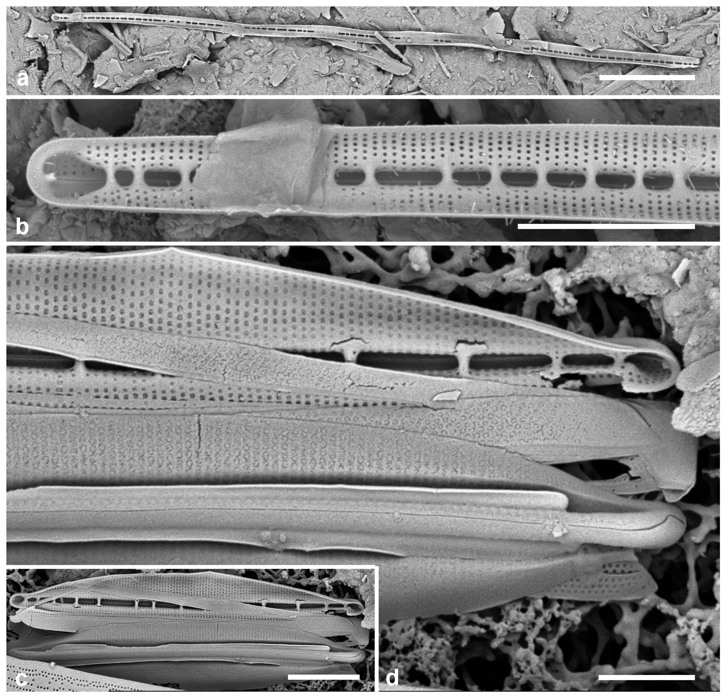
3.170. Homoeocladia volvendirostrata (Ashworth, Dᶏbek & Witkowski) Lobban and Ashworth 2022 [197]—Figure 57c,d
Yap samples: Y37-8
Dimensions: Length 13–17 µm, width 1.7–3 µm, striae 52 in 10 µm, fibulae 7–11 in 10 µm
3.171. Nitzschia frustulum (Kützing) Grunow 1880 in [68]
Dimensions: Length 17 µm, width 2.4 µm, striae 20–22 in 10 µm, fibulae 10 in 10 µm
3.172. Nitzschia longissima (Brébisson ex Kützing) Ralfs 1861 in [50]—Figure 58a–c
Yap samples: Y25H-2, Y26B, Y37-8, Y41-7
Dimensions: Length 204 µm, striae 34 in 10 µm.
3.173. Nitzschia maiae Lobban, Ashworth, Calaor & E.C.Theriot 2019 [157]—Figure 58d,e
Yap samples: Y36-1, Y36-5. Y41-7, Y41-8.
Dimensions: Length 33 µm, width 4 µm, striae 37 in 10 µm, fibulae 17 in 10 µm
Diagnostics: Very small conopea only at the apices, lacking a valve depression, and easily distinguished from Homoeocladia spp. by the high fibula density.
3.174. Nitzschia marginulata var. didyma Grunow 1880 in [200]—Figure 58f
Yap samples: Y34C, Y34H, Y34F, Y36-2, Y36-5
Dimensions: Length 27–36 µm, width 9–10 µm, striae 28 in 10 µm, fibulae 16 in 10 µm
Nitzschia spp. (a–c) Nitzschia longissima, SEM. (a,b) Whole valve, external view, with detail of central portion. (c) Internal aspect near central area showing uninterrupted striae and character of fibulae. (d,e) N. maiae, SEM and LM of frustule. (f) N. marginulata var. didyma frustule SEM. Scale bars: (a) = 25 µm, (b–f) = 10 µm.
Figure 58.
Nitzschia spp. (a–c) Nitzschia longissima, SEM. (a,b) Whole valve, external view, with detail of central portion. (c) Internal aspect near central area showing uninterrupted striae and character of fibulae. (d,e) N. maiae, SEM and LM of frustule. (f) N. marginulata var. didyma frustule SEM. Scale bars: (a) = 25 µm, (b–f) = 10 µm.

3.175. Nitzschia obtusa f. parva Hustedt 1921 in [163]
Dimensions: Length 35–40 µm, width 5–7 µm, striae 30–35 in 10 µm
3.176. Nitzschia pseudohybridopsis Lobban, sp. nov.—Figure 59 and Figure 60
Diagnosis: Nitzschiae dubiae differing from N. pseudohybrida in the sparser fibulae.
Nitzschia pseudohybridopsis, sp. nov. (a–c) Range of size in LM, (c) = holotype. (d) Frustule in girdle view, SEM, showing ‘papillae’ on valvocopula. (e) Valve in valve view. Scale bars: (a–c) = 10 µm, (d,e) = 5 µm.
Figure 59.
Nitzschia pseudohybridopsis, sp. nov. (a–c) Range of size in LM, (c) = holotype. (d) Frustule in girdle view, SEM, showing ‘papillae’ on valvocopula. (e) Valve in valve view. Scale bars: (a–c) = 10 µm, (d,e) = 5 µm.
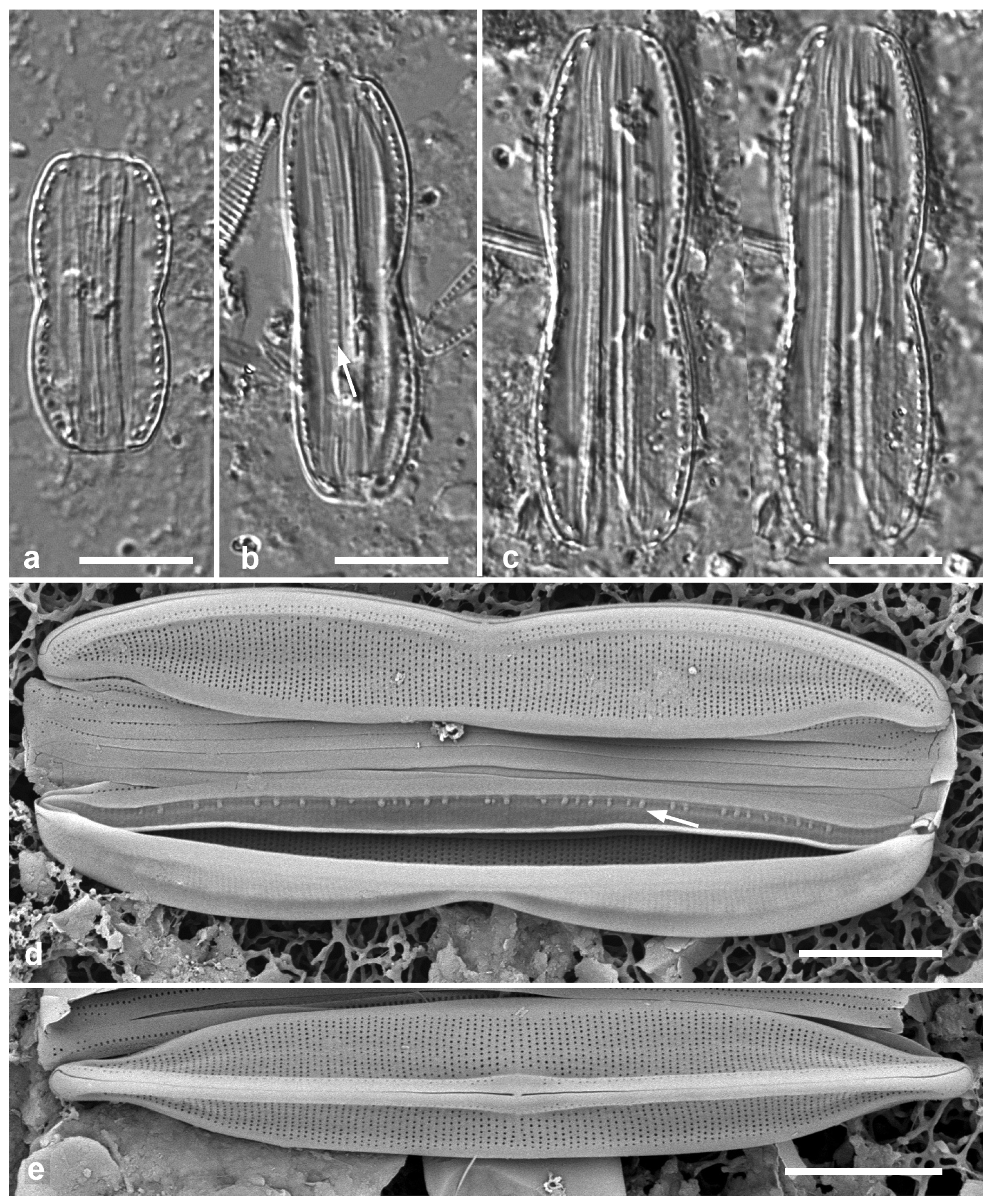
Type locality: YAP STATE: Maap Island, Wanead, 9°36′15″ N, 138°11′0″ E; estuarine, intertidal rocks from mouth of stream, sample Y16B. Coll. C.S. Lobban & M. Schefter, 21 September 1988. Plentiful in this sample.
Nitzschia pseudohybridopsis, sp. nov., cont. (a,b) SEM of frustules showing details of the valvocopula (VC) with small pars exterior (pe) and larger papillate pars interior (pi), (b) also showing part of the internal keel with central nodule. Scale bars: 5 µm.
Figure 60.
Nitzschia pseudohybridopsis, sp. nov., cont. (a,b) SEM of frustules showing details of the valvocopula (VC) with small pars exterior (pe) and larger papillate pars interior (pi), (b) also showing part of the internal keel with central nodule. Scale bars: 5 µm.

Etymology: Named for its resemblance to N. pseudohybrida.
Additional materials: Y26C
3.177. Nitzschia ventricosa J.L. Palmer 1873 in [201]—Figure 61a
Yap samples: Y26B
Dimensions (from Guam records): Length 150 µm, width 10 µm, striae 34 in 10 µm, fibulae and costae in central inflation 7–10 in 10 µm
Comments: Only one fragment observed in Yap samples.
3.178. Psammodictyon constrictum (W. Gregory) D.G. Mann 1990 in [52]—Figure 61b
Yap samples: Y25H-1, Y25H-2, Y37-7, Y37-8, Y36-1
Dimensions: Length 22 µm, width 9 µm, striae 16 in 10 µm, fibulae 16 in 10 µm
3.179. Psammodictyon panduriforme (W. Gregory) D.G. Mann 1990 in [52]—Figure 61d,e
Yap samples: Y25H-1, Y26C, Y37-7, Y36-1
Dimensions: Length 90–100 µm, width 25 µm, striae 16–18 in 10 µm, fibulae 8 in 10 µm
3.180. Psammodictyon pustulatum (Voigt ex Meister) Lobban 2015 [13]—Figure 61c
Yap samples: Y18E, Y36-4, Y37-8
Dimensions: Length 27–32 µm, width 15–17 µm, striae 22 in 10 µm
Diagnostics: This species is similar in size and areola pattern to P. constrictum but differs in the relief of the valve surface and in the presence of a longitudinal break across the striae.
3.181. Tryblionella granulata (Grunow) D.G.Mann 1990 in [52]—Figure 62a
Yap samples: Y7I, Y34A
Dimensions: Length 16–24 µm, width 6–9 µm, striae 7.5 in 10 µm
(a) Nitzschia ventricosa, valve fragment, LM. (b) Psammodictyon constrictum frustule in SEM. (c) Psammodictyon pustulatum frustule in SEM. (d,e) Psammodictyon panduriforme, SEM. (d) Frustule showing exterior valve face. (e) Internal valve face. Scale bars: (a,d,e) = 10 µm, (b,c) = 5 µm.
Figure 61.
(a) Nitzschia ventricosa, valve fragment, LM. (b) Psammodictyon constrictum frustule in SEM. (c) Psammodictyon pustulatum frustule in SEM. (d,e) Psammodictyon panduriforme, SEM. (d) Frustule showing exterior valve face. (e) Internal valve face. Scale bars: (a,d,e) = 10 µm, (b,c) = 5 µm.
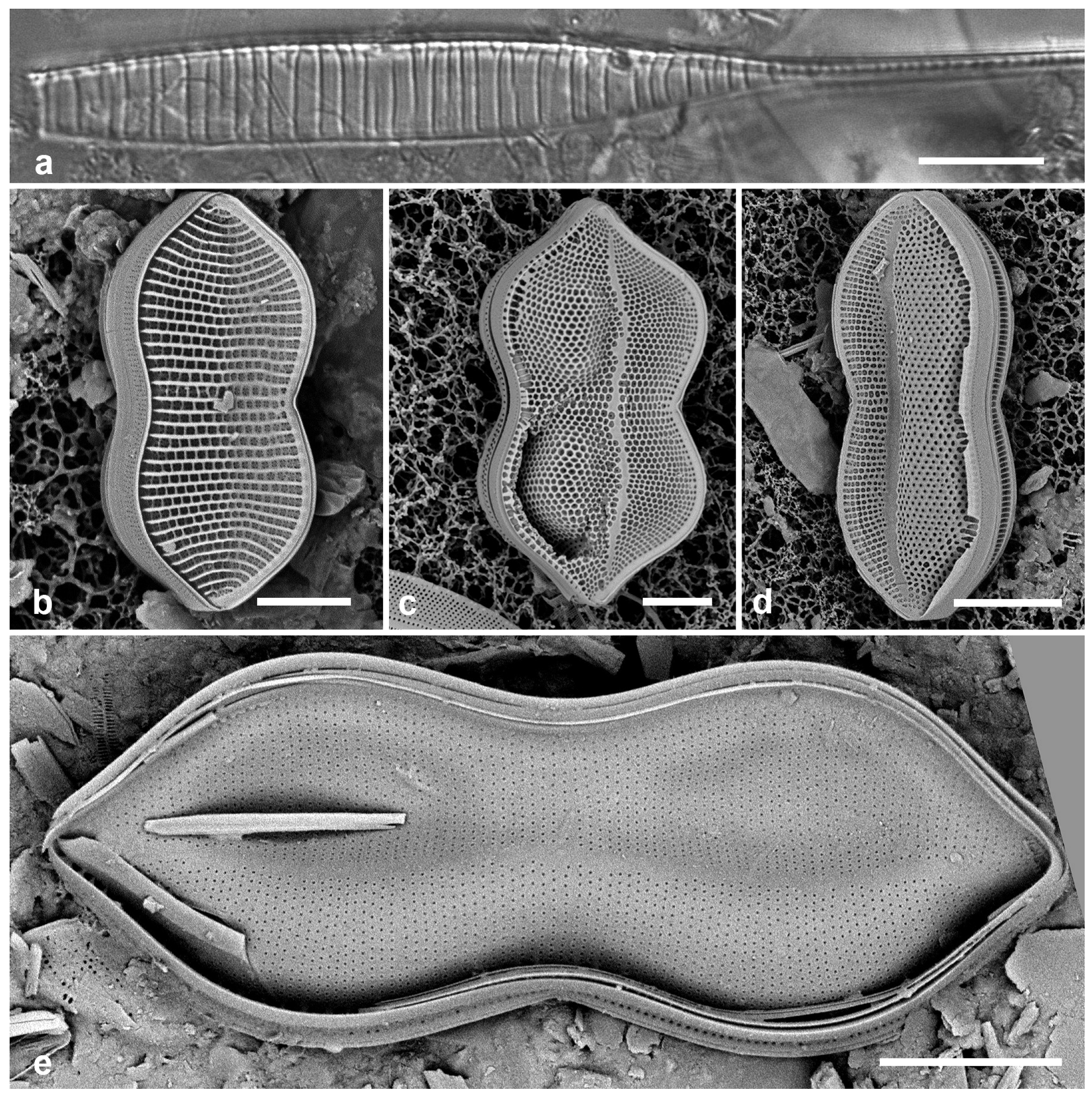
3.182. Entomoneis yudinii Prelosky & Lobban, sp. nov.—Figure 62b–g
Diagnosis: Moderately silicified compared to congeners with prominent areolae in uniseriate striae.
(a) Tryblionella granulata, frustule, SEM, Yap voucher, courtesy of Nelson Navarro. (b–g) Entomoneis yudinii, sp. nov. (b,c) Valves in valve view, LM; (c) = holotype. (d) Valve in valve view showing overall shape and features of exterior. (e) Detail of valve showing depressed area with infilled areolae and the faint asymmetrical stauros (arrows). (f) Detail of valve in (d) showing outer coverings of areolae and a grooved rib alongside the raphe slit (arrow). (g) Interior showing smaller inner foramina, also occluded, and part of the keel canal with fibulae (arrow). Scale bars: (b–d) = 10 µm, (a,e,g) = 5 µm, (f) = 2 µm.
Figure 62.
(a) Tryblionella granulata, frustule, SEM, Yap voucher, courtesy of Nelson Navarro. (b–g) Entomoneis yudinii, sp. nov. (b,c) Valves in valve view, LM; (c) = holotype. (d) Valve in valve view showing overall shape and features of exterior. (e) Detail of valve showing depressed area with infilled areolae and the faint asymmetrical stauros (arrows). (f) Detail of valve in (d) showing outer coverings of areolae and a grooved rib alongside the raphe slit (arrow). (g) Interior showing smaller inner foramina, also occluded, and part of the keel canal with fibulae (arrow). Scale bars: (b–d) = 10 µm, (a,e,g) = 5 µm, (f) = 2 µm.
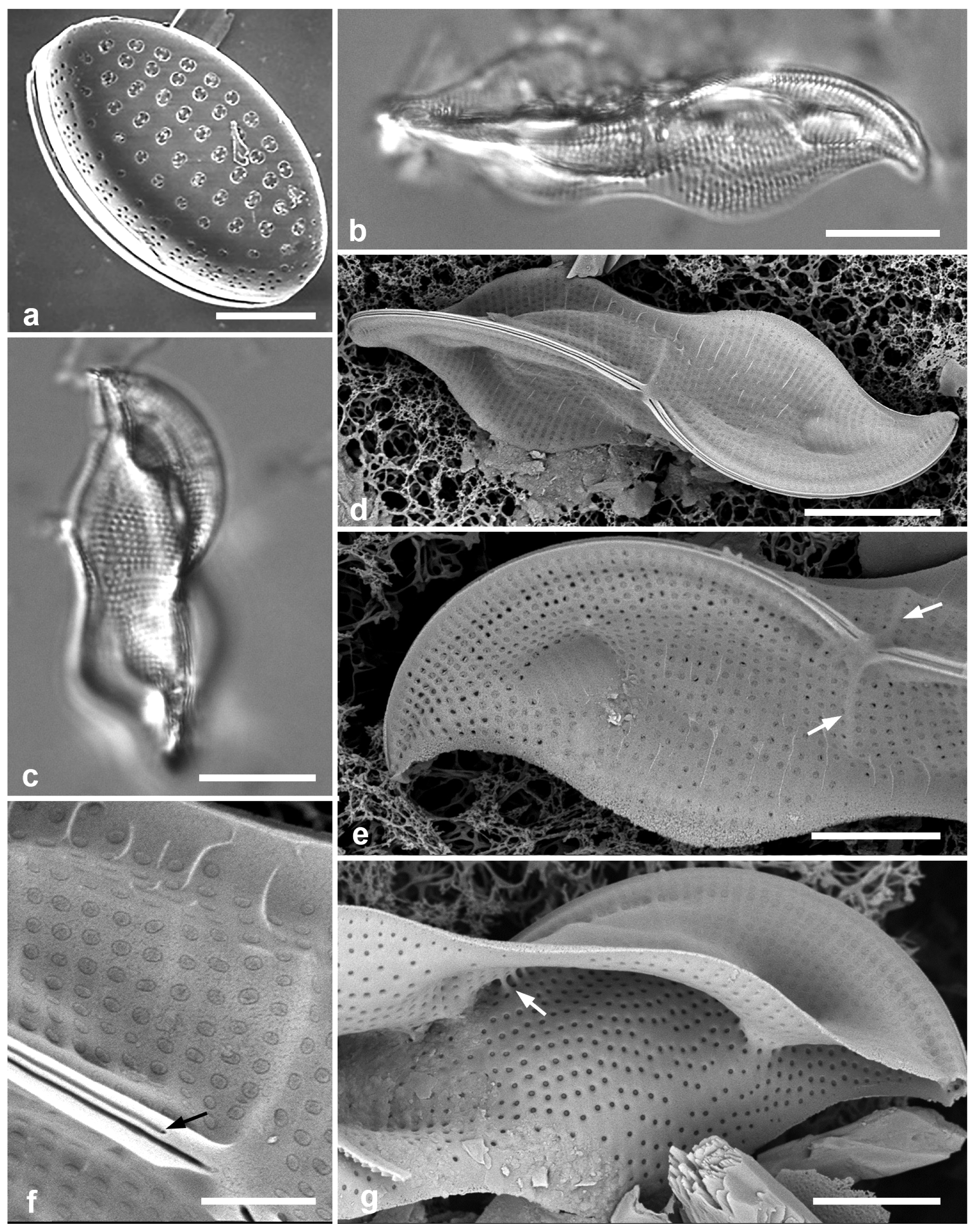
Type locality: Yap State, Yap Island, Weloy Municipality, Maa’ Mangrove, approx: 9°32′25.62″ N, 138° 5′14.45″ E, adjacent to Nimpal Marine Protected Area, sample Y34A, subtidal mud from Sonneratia alba (white mangrove) pneumorrhiza, ca. 20 cm from bottom, and likely in the salt wedge. Coll. C.S. Lobban, M. Schefter & T. Gorong, 28 May 2014.
Etymology: Named in honor of Lee Yudin, former Dean of the College of Natural and Applied Sciences, University of Guam, for his support of the Microscopy Teaching and Research Laboratory over many years and for mentoring Prelosky.
Additional records: YAP: Y34B
Rhopalodiaceae (Karsten) Topachevs’kyj & Oksiyuk
3.183. Epithemia guettingeri (K. Krammer) Lobban & Joon S. Park in [15]—Figure 63a–c
Additional Yap samples: Y26C, Y29C, Y34A, Y34B, Y36-5, Y39A
Dimensions: Length 19–54 µm, width 7–11 µm, striae ca. 30–40 in 10 µm
3.184. Epithemia muscula Kützing 1844 [37]—Figure 63d,e
Yap samples: Y37-8
Dimensions: Length 12–30 µm, width 21 µm, areolae 21 in 10 µm
Epithemia spp. in SEM. (a–c) E. guettingeri. (a) Valve view of ventral surface, showing axial “costa” formed by narrow wave in surface (arrow), note areolae in the groove (arrowhead). Stria density about 30 along keel. (b) Small specimen, ca. 40 striae, axial costa not so clear. (c) Interior aspect of valve showing transverse costae and part of keel canal (Palau specimen PW(2022)1A-5). (d,e) Epithemia muscula, SEM. (d) Frustule in oblique dorsal view, showing narrow dorsal surface (arrow). (e) Interior aspect of valve showing numerous transverse costae and large cribrate areolae. Scale bars = 5 µm.
Figure 63.
Epithemia spp. in SEM. (a–c) E. guettingeri. (a) Valve view of ventral surface, showing axial “costa” formed by narrow wave in surface (arrow), note areolae in the groove (arrowhead). Stria density about 30 along keel. (b) Small specimen, ca. 40 striae, axial costa not so clear. (c) Interior aspect of valve showing transverse costae and part of keel canal (Palau specimen PW(2022)1A-5). (d,e) Epithemia muscula, SEM. (d) Frustule in oblique dorsal view, showing narrow dorsal surface (arrow). (e) Interior aspect of valve showing numerous transverse costae and large cribrate areolae. Scale bars = 5 µm.
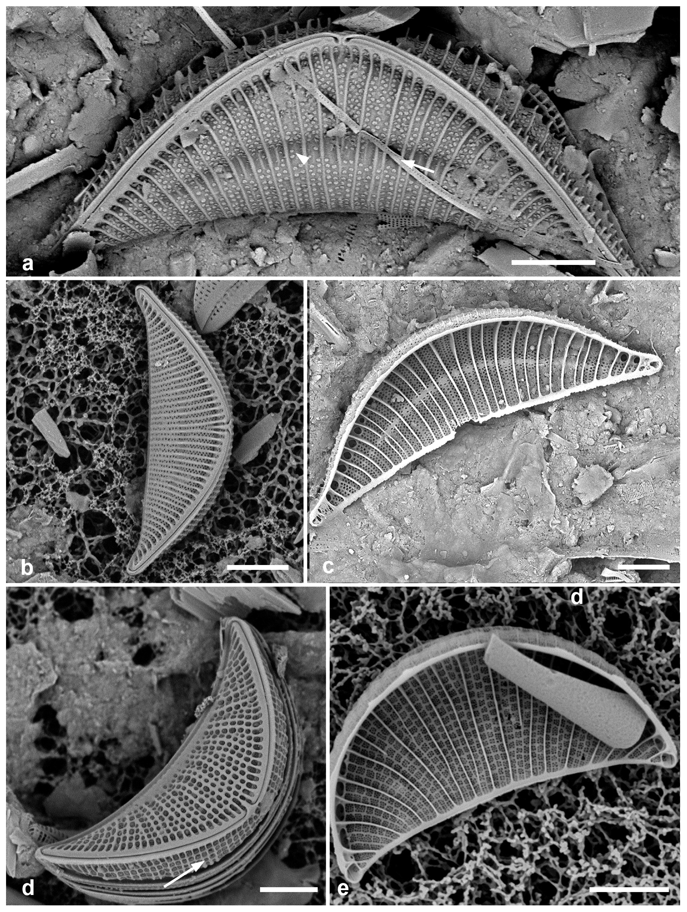
3.185. Protokeelia cholnokyi (Giffen) F.E.Round & P.W.Basson 1995 [207]—Figure 64a,b
Yap samples: Y26B, Y26C
Dimensions: Length 5–17 µm, width 4–7 µm, biseriate striae ca. in 10 µm
(a,b) Protokeelia cholnokyi valve in LM and frustule in SEM. (c) Auricula intermedia. (d) Campylodiscus giffenii valve, SEM. (e) Campylodiscus humilis valve, LM. Scale bars: (a,c,e) = 10 µm, (b,d) = 5 µm.
Figure 64.
(a,b) Protokeelia cholnokyi valve in LM and frustule in SEM. (c) Auricula intermedia. (d) Campylodiscus giffenii valve, SEM. (e) Campylodiscus humilis valve, LM. Scale bars: (a,c,e) = 10 µm, (b,d) = 5 µm.
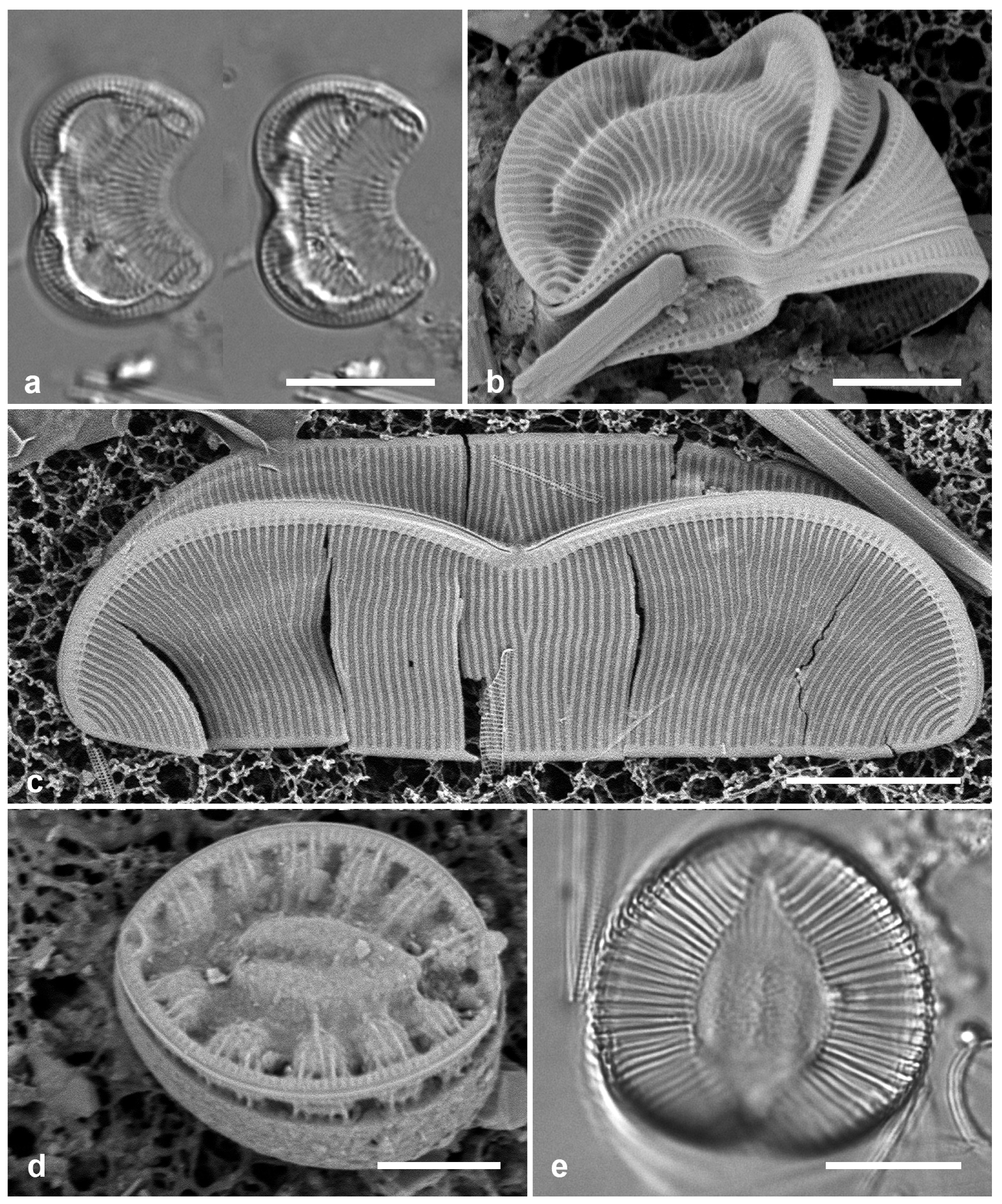
3.186. Auricula complexa (W. Gregory) Cleve 1894 [162]
Dimensions: Length 45 µm, striae 18–20 in 10 µm
3.187. Auricula flabelliformis M.Voigt 1960 [208]
Additional Yap records: Y26C
Dimensions: Length 92–95 μm, width 69 μm, striae ca. 19 in 10 μm; fibulae ca. 8 in 10 μm.
Comments: There is another large Auricula in the samples not yet identified.
3.188. Auricula intermedia (Lewis) Cleve 1894 [162]—Figure 64c
Yap samples: Y26B, Y26C, Y41-7
3.189. Campylodiscus brightwellii Grunow 1862 [158]
Yap samples: Y25H-1, Y25H-2, Y42-1
Dimensions: Diam. 25–30 µm
3.190. Campylodiscus giffenii Lobban & JoonS.Park 2022 in [16]—Figure 64d
Synonyms: Surirella scalaris Giffen
Campylodiscus scalaris (Giffen) Lobban & JoonS. Park
Yap samples: Y34B, Y41-8
Dimensions: Length 10–13 µm, width 10 µm
3.191. Campylodiscus humilis Greville 1865 [4]—Figure 64e
Yap samples: Y37-8
Dimensions: Diam. 23–29 μm
Comments: This and the following species probably belong in Coronia but have not been transferred.
3.192. Campylodiscus imperialis Greville 1860 [209]
Dimensions: Diam. 40–50 μm
3.193. Campylodiscus neofastuosus Ruck & Nakov 2016 in [206]—Figure 66a
Yap samples: Y25H-2, Y37-8, Y36-1, Y41-7, -8
Dimensions: Length 39–90 μm, width 28–71 µm
3.194. Campylodiscus ralfsii W.Smith 1853 [210]
Dimensions: Diam. 60–70 µm
3.195. Campylodiscus tatreauae Prelosky & Lobban, sp. nov.—Figure 65
Diagnosis: Differing from C. neofastuosus in the aqueduct-like rim (raphe-keel), more prominent, barred fenestrae, and silica fins on the infundibular costae.
Type locality: Yap State, Yap Island, Weloy Municipality, Maa’ Mangrove, approx: 9°32′25.62″ N, 138° 5′14.45″ E, adjacent to Nimpal Marine Protected Area, sample Y34A, subtidal mud from Sonneratia alba (white mangrove) pneumorrhiza, ca. 20 cm from bottom, and likely in the salt wedge. Coll. C.S. Lobban, M. Schefter & T. Gorong, 28 May 2014
Etymology: Named in honor of Prelosky’s high-school science teacher and mentor, Linda Tatreau, whose Marine Mania program introduced many students to marine biology.
Additional material: Y34B, Y34C; PALAU: PW(2021)1-1, PW(2021)4-7, PW(2022)1-1, PW(2022)1A-5; GUAM: GU58G-1.
Campylodiscus tatreauae sp. nov. Yap (a–c) and Palau (d–g). (a) Holotype in LM showing barred fenestrae (arrow). (b) Valve exterior showing aqueduct-like rim (double-headed arrow), silica flaps, and barred fenestrae (arrow). (c,d) Details of margin with barred fenestrae (arrows) (c) in LM from another specimen on same slide, (d) in SEM (PW(2022)1A-5). (e) Valve interior. (f) Detail of valve interior (same specimen). (g) Oblique external view of valve showing rim (double-headed arrow) and silica flaps. Scale bars: (a–c,e) = 10 µm, (d,f,g) = 5 µm.
Figure 65.
Campylodiscus tatreauae sp. nov. Yap (a–c) and Palau (d–g). (a) Holotype in LM showing barred fenestrae (arrow). (b) Valve exterior showing aqueduct-like rim (double-headed arrow), silica flaps, and barred fenestrae (arrow). (c,d) Details of margin with barred fenestrae (arrows) (c) in LM from another specimen on same slide, (d) in SEM (PW(2022)1A-5). (e) Valve interior. (f) Detail of valve interior (same specimen). (g) Oblique external view of valve showing rim (double-headed arrow) and silica flaps. Scale bars: (a–c,e) = 10 µm, (d,f,g) = 5 µm.
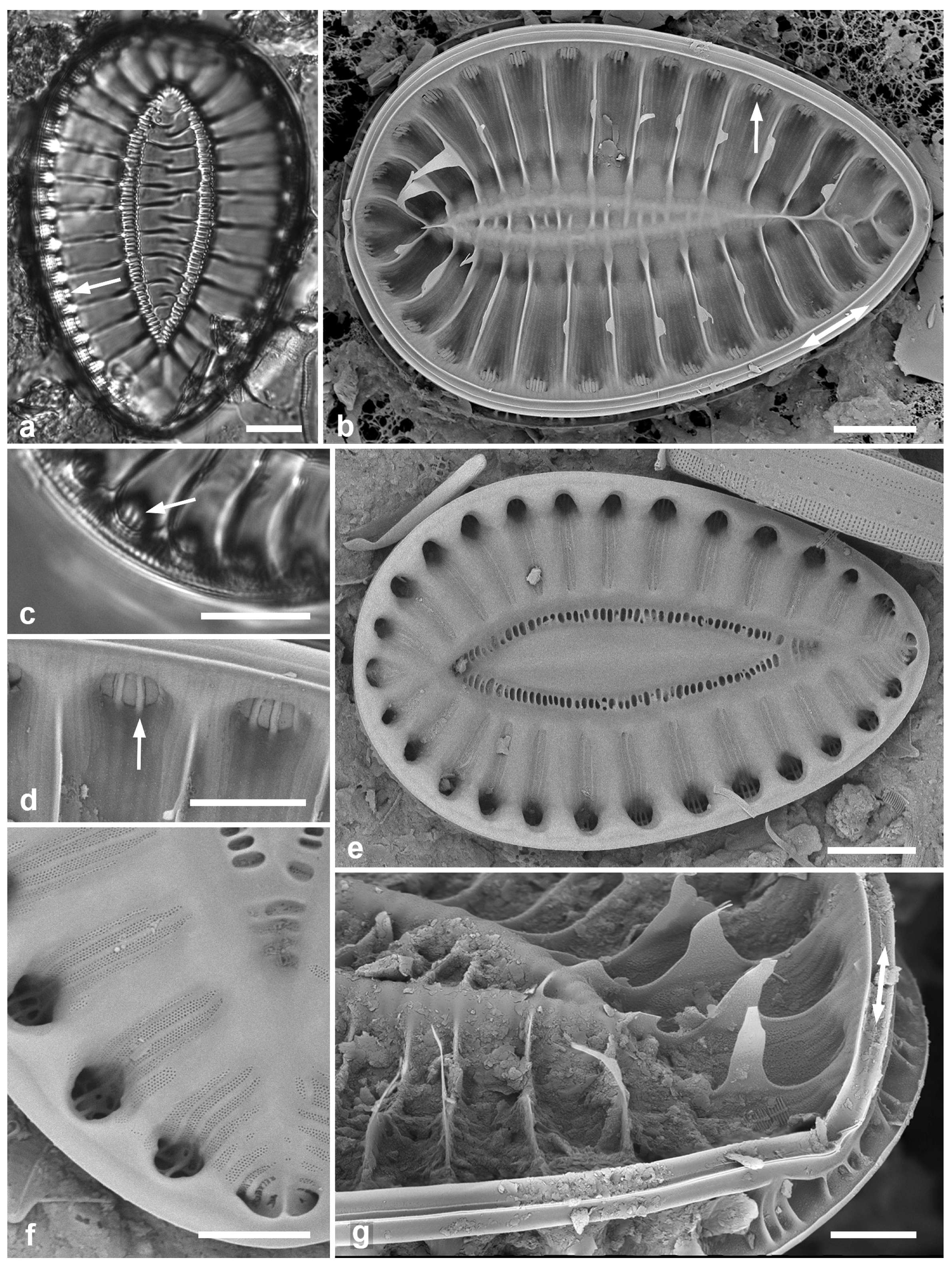
(a) Campylodiscus neofastuosus, valve in SEM, showing small fenestrae and “pie-crust” rim (arrows). (b) Coronia ambigua, LM. (c) Coronia decora var. decora, LM, frustule showing two axes at right angles. (d) Coronia decora var. pinnata valve, LM. (e,f) Hydrosilicon mitra, SEM. (e) Yap voucher. (f) Detail of middle of valve interior, Guam specimen, showing striae converging on transverse and longitudinal ribs (see also Figure 67a,b). Scale bars: (a–e) = 10 µm, (f) = 5 µm.
(a) Campylodiscus neofastuosus, valve in SEM, showing small fenestrae and “pie-crust” rim (arrows). (b) Coronia ambigua, LM. (c) Coronia decora var. decora, LM, frustule showing two axes at right angles. (d) Coronia decora var. pinnata valve, LM. (e,f) Hydrosilicon mitra, SEM. (e) Yap voucher. (f) Detail of middle of valve interior, Guam specimen, showing striae converging on transverse and longitudinal ribs (see also Figure 67a,b). Scale bars: (a–e) = 10 µm, (f) = 5 µm.
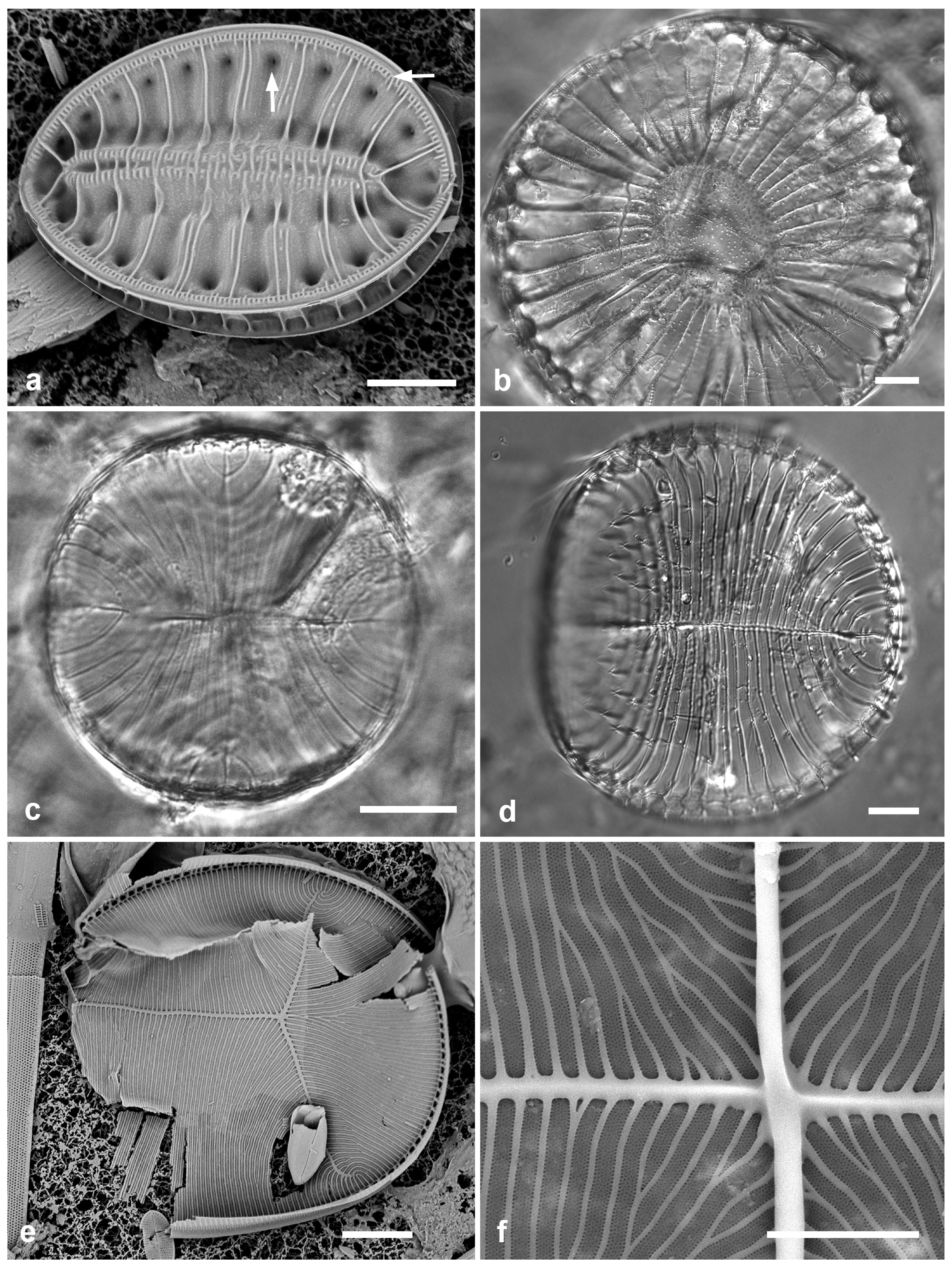
3.196. Campylodiscus wallichianus Greville 1863 [212]
Dimensions: Diam. 75–90 μm
3.197. Coronia ambigua (Greville) Ruck & Guiry 2016 [213]—Figure 66b
Yap samples: Y26B
Dimensions: Diam. 104 µm
3.198. Coronia decora (Brébisson) Ruck & Guiry 2016 [213]—Figure 66c
Yap samples: Y25H-2, Y26B, Y26C
Dimensions: Diam. 39–41 µm
3.199. Coronia decora var. pinnata (Peragallo) Lobban & J.S. Park in [15]—Figure 66d
Yap samples: Y26B
Dimensions: Diam. 72–80 µm
3.200. Hydrosilicon mitra Brun 1891 [214]—Figure 66e,f and Figure 67
Yap samples: Y37-8
Dimensions: Length ca. 100 μm, width 50 μm, striae 17 in 10 μm
3.201. Petrodictyon gemma (Ehrenberg) D.G. Mann 1990 in Round [52]—Figure 68a–c
Yap samples: Y34A
Dimensions: Length 75–76 µm, width 43 µm; striae 20 in 10 µm, areolae ca. 24 in 10 µm
3.202. Petrodictyon patrimonii (Sterrenburg) Sterrenburg 2001 [215]—Figure 68d–f
(a,b) Hydrosilicon mitra, Guam specimen, showing entire specimen, valve internal view, and higher magnification of constriction where the two raphe endings meet in the nodule and from whence the striae radiate towards the transverse and longitudinal costae. Scale bars: (a) = 10 µm, (b) = 5 µm.
Figure 67.
(a,b) Hydrosilicon mitra, Guam specimen, showing entire specimen, valve internal view, and higher magnification of constriction where the two raphe endings meet in the nodule and from whence the striae radiate towards the transverse and longitudinal costae. Scale bars: (a) = 10 µm, (b) = 5 µm.

Additional Yap samples: Y26B
Dimensions: Length 77–84 μm, width 15–29 μm, striae 30 in 10 μm
Comments: See P. gemma, above.
Petrodictyon. (a–c) Petrodictyon gemma. (a) LM showing visible striae and areolae. (b) Half of external valve view, SEM. (c) Half of internal view, SEM, inset showing 2–3 portulae between fibulae. (d–f) Petrodictyon patrimonii. Fig. (d) LM showing striae not resolved. (e) Internal view, SEM, inset showing 3–6 portulae between fibulae. (f) Frustule (broken open), showing depth of mantle. Scale bars = 10 µm.
Figure 68.
Petrodictyon. (a–c) Petrodictyon gemma. (a) LM showing visible striae and areolae. (b) Half of external valve view, SEM. (c) Half of internal view, SEM, inset showing 2–3 portulae between fibulae. (d–f) Petrodictyon patrimonii. Fig. (d) LM showing striae not resolved. (e) Internal view, SEM, inset showing 3–6 portulae between fibulae. (f) Frustule (broken open), showing depth of mantle. Scale bars = 10 µm.
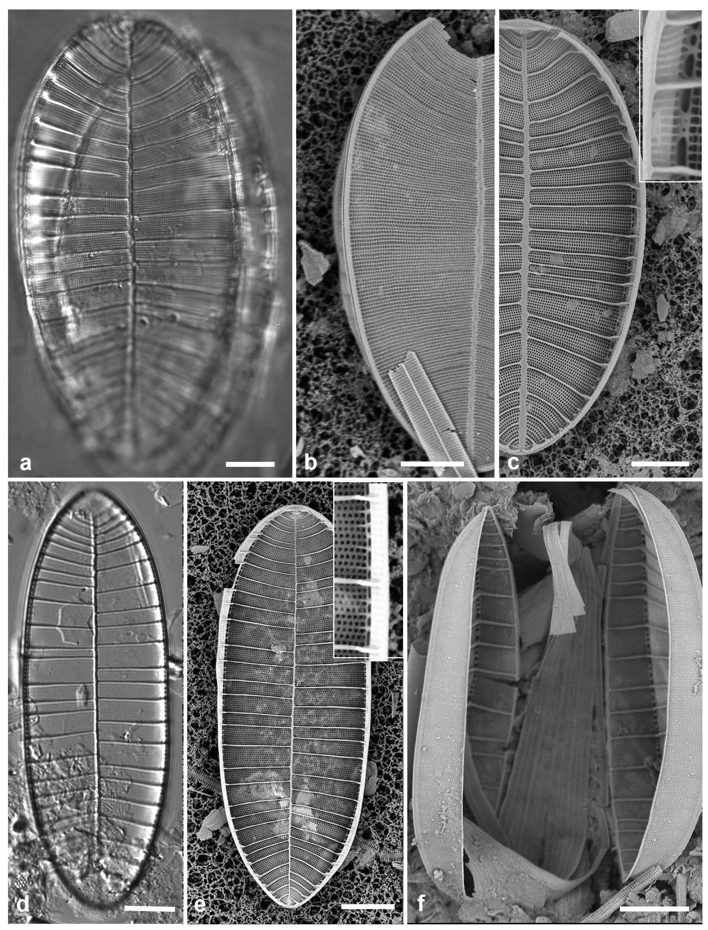
Source link
Christopher S. Lobban www.mdpi.com

